Emergency War Surgery (2004) - page 5
Damage Control Surgery
occurs before the site can be identified; the liver is packed
again; the patient is resuscitated again; and the entire cycle
is repeated.
ο Definitive therapeutic packing is based on three basic
principles.
♦ Pressure stops bleeding.
♦ Pressure vectors should recreate tissue planes (attempt
to recreate the pressure vectors created by the capsule
of a solid organ or fill the space of that organ, not
random pack placement).
♦ Tissue viability must be preserved.
ο 6-12 laparotomy pads are the best commonly available
packing material.
ο An intervening layer, such as a bowel bag, sterile drape,
absorbable mesh, or omentum, can be placed between
packs and the tissue to aid in easy pack removal at
relaparotomy.
Abdominal Closure.
ο
Leave the fascia open.
ο
Vacuum pack (preferred technique — easy, keeps patient
dry, allows for expansion).
♦ With fascia open, place fully plastic-covered (bowel bag,
X-ray cassette bag, Ioban drape) sterile operating room
(OR) towel circumferentially under the fascia to cover
the viscera. Place a small number of central perforations
to allow fluid to egress to the drains.
♦ Place closed-suction drains (Jackson-Pratt, modified
Foley, small chest tube) above the plastic at the level of
the subcutaneous tissue brought out through separate
stab wounds or the inferior portion of the wound.
♦ Place lap sponges to fill in the wound.
♦ Cover the entire wound with a large Ioban drape.
♦ Place drains on low suction and secure to the skin.
ο
A silastic sheet or 3-liter IV bag, sewn to the skin or fascia,
can accomplish abdominal closure in virtually every
instance.
ο
Skin closure is not recommended, but may be quickly
accomplished with skin staples, towel clips (reliably
stronger), or running monofilament suture.
12.5
Emergency War Surgery
Skin closure may lead to abdominal compartment
syndrome.
Thoracic injuries
The goal of abbreviated thoracotomy is to stop the bleeding
and restore a survivable physiology; contamination is
usually not a problem.
In the exsanguinating patient, formal lung resection gives
way to using large staplers in a nonanatomic wedge resection
to rapidly achieve hemostasis and control of air leaks.
In pulmonary tractotomy, the lung bridging the wound tract
is opened between long clamps or with a linear stapler. The
tract can thus be directly inspected, bleeding points selectively
ligated, and air leaks controlled.
Vascular injuries can be treated with intraluminal shunts or
Fogarty balloons to achieve distal control in inaccessible areas.
Tracheal injury can be treated with airway control placed
through the site of injury.
Extensive bronchial repairs are not feasible in the patient in
extremis; therefore, rapid resection of the affected lobe or lung
would be best.
When dealing with esophageal injury, diversion, and wide
drainage, not definitive repair, is the best course of action.
A single en masse suture closure of the chest wall is best
because wound closure of skin with towel clips may result in
significant blood loss from the musculature.
Phase 2: Critical Care Considerations
Physiologic support in the post-op TASC patient is
paramount to survival.
ο Core rewarming: warmed resuscitative fluids, blankets,
ventilator air, and environment, or commercially available
products such as Bair Hugger, Chill Buster.
ο Reversal of acidosis: appropriate/aggressive resuscitation
with crystalloid, colloid, and blood products.
ο Reversal of coagulopathy: at many locations, only ultra-
fresh whole blood is available to correct coagulopathy.
Abdominal compartment syndrome.
12.6
Damage Control Surgery
ο
Abdominal compartment syndrome is a condition in which
increased intra-abdominal pressure adversely affects the
circulation and threatens the function and viability of
the viscera.
ο
Measurement is performed using urinary bladder pressure
(normal = zero).
♦ Several methods are available for performing bladder
pressure.
◊ Place 100-150 cc of sterile saline in the bladder and
clamp the foley.
◊ Access the needle port on the catheter and attach to a
pressure monitor (central venous pressure transducer).
◊ Access the needle port and create a column of water
via plain IV tubing held vertically or use of the
pressure gauge from a lumbar puncture kit.
◊ If there is no needle port, clamp the foley proximal
and the distal end of regular IV tubing in the usual
drainage end of the catheter until firmly in place.
♦ Measurement of bladder pressure is a good variable to
test and follow; however, intervention for abdominal
compartment syndrome (ACS) should occur when
suspected or clinically indicated.
ο
Occurs in abdominal trauma accompanied by visceral
swelling, hematoma, or abdominal pack use.
ο
Physiology of abdominal compartment syndrome.
♦ Cardiac output and venous return are decreased.
♦ Reduction in blood flow to liver, intestines, and kidneys
can result in anuria.
♦ The two hemidiaphragms push upward, decreasing
thoracic volume, and compliance leading to elevated
peak airway pressures.
♦ Central venous, pulmonary capillary wedge, and right
atrial pressures increase with intra-abdominal pressure
(can lead to false PA catheter pressures).
♦ PO2 is decreased due to increases in airway pressures
and ventilation/perfusion abnormalities that worsen
with positive end-expiratory pressure (PEEP).
12.7
Emergency War Surgery
Abdominal Pressure Degree of Elevation Clinical Effect
10-20 mm Hg
Mild
Insignificant
20-40 mm Hg
Moderate
Oliguria and organ dysfunction
>40 mm Hg
Severe
Requires immediate attention
Phase 3: Planned Reoperation
Packs should be left in place until the patient’s hemodynamics
are stable and all major sites of hemorrhage have had time to
clot.
Reoperation should be scheduled when the probability of
achieving definitive organ repair and complete fascial closure
are highest.
Timing must coincide with reversal of hypotension, acidosis,
hypothermia, and coagulopathy. Typically occurs 24-48 hours
following the primary insult when brisk diuresis, negative
fluid balance, diminishing abdominal girth, and decreasing
peripheral edema indicate reduction in visceral and parietal
edema.
This surgery may occur at the next echelon of care.
ο Stratevac should be weighed carefully because transit
operative care is minimal. Surgical expertise is generally not
available and transit times are often greater than 24 hours.
Timing can, however, be dictated by other pressing clinical
concerns such as abdominal compartment syndrome, limb
ischemia, and suboptimal control of spillage at primary
operation.
In cases of a packed and drained duodenum, pancreas, kidney
or bladder, or liver injuries with gross bowel contamination,
packs should be retrieved within 36-48 hours.
It is sometimes necessary to perform this type of operation
at the bedside, as the patient’s cardiopulmonary status does
not allow a trip to the OR.
Conduct of Relaparotomy
It is to be presumed that injuries were missed.
A complete laparotomy must be performed in search of
missed injury.
12.8
Damage Control Surgery
The surgeon must exercise caution and sound judgment
before performing full reconstruction of the GI tract because
the patient is typically still critically ill and catabolic, making
the patient less likely to heal anastomoses and even less likely
to tolerate a leak or uncontrolled fistula.
Feeding tube placement, either transabdominal or naso-
enteric, should be placed at this time.
Repacking may be re-employed if other measures fail to
control hemorrhage.
An abdominal film should be obtained to insure all packs
have been removed from the abdomen. Sponge counts should
be considered unreliable in this situation.
Unplanned Reexploration.
ο Emergent, unplanned reexploration should be performed
in any:
♦ Normothermic patient with unabated bleeding (> 2
units of PRBCs/h).
♦ Patient who develops severe intra-abdominal compart-
ment syndrome.
♦ Patient requiring postoperative transfusion of > 10 units
of PRBCs.
♦ Patient with persistent lactic acidosis.
Austere Field and Military Surgical Considerations
Due to severe physiological insult, the typical civilian damage
control patient requires 2 surgeons and 1 nurse, at a minimum,
at the bedside for the first 6 hours. An example of the magni-
tude of the ICU problem that may be encountered is a casualty
who requires a pulmonary artery catheter, 3 operations, 33
units of PRBCs, and an ICU stay of 23 days. In mass casualty
scenarios, this patient would likely be triaged as expectant.
Tactical Abbreviated Surgical Control philosophy allows
the surgeon to apply damage control techniques when the
limitations of reserve exist outside the patient, in the
tactical environment, not just to patients about to exhaust
their physiological reserve (classic damage control
scenario).
12.9
Emergency War Surgery
Summary Points
Damage control is not a procedure of last resort. The
consideration of damage control techniques should be made
at the initiation of operative intervention in any multitrauma
casualty and reconsidered during any case where extensive
injuries are involved.
Consider damage control in patients with severe liver injury,
combined vascular and hollow viscus injuries, multiple sites
of hemorrhage, diminished physiological reserve, and com-
binations of severe injury involving multiple organ systems
(eg, CNS, orthopedic, vascular, or thoracoabdominal).
Consider damage control early, ISS > 35, pH < 7.2, temp-
erature < 34°C, shock, or coagulopathy.
Avoid: hypothermia, ‘pack and peek’, tight abdominal
closure, abdominal compartment syndrome, delayed
relaparotomy for surgical bleeding, and getting stuck in a
conventional thinking mode.
Think: vascular shunts/fasciotomy, abdominal packing,
external fixators, angiographic embolization (when avail-
able), temporary closure, nonanatomic resections, and missed
injury.
Plan sequence of operation in stable patients to allow for use
of damage control if instability develops.
12.10
Chapter 13
Face and Neck Injuries
Introduction
Immediate recognition and appropriate management of
airway compromise is critical to survival.
Face and neck injuries can be the most difficult-to-manage
wounds encountered by health care providers in the combat
zone. Focusing on ABC priorities is vital.
During airway control, maintain cervical spine
immobilization in bluntly injured patients. (Unstable C-spine
injury is very rare in neurologically intact penetrating face
and neck wounds.)
Bleeding should be initially controlled with direct pressure.
If bleeding cannot be controlled, immediate operative
intervention is necessary.
Complete assessment of remaining injuries (fractures,
lacerations, esophageal injury, ocular injuries).
Immediate Management of Facial Injuries
Airway.
ο Airway distress due to upper airway obstruction above
the vocal cords is generally marked by inspiratory stridor:
♦ Blood or edema resulting from the injury.
♦ Tongue may obstruct the airway in a patient with a
mandible fracture.
♦ A fractured, free-floating maxilla can fall back,
obstructing the airway.
♦ Displaced tooth fragments may also become foreign
bodies.
ο Maneuvers to relieve upper airway obstruction:
13.1
Emergency War Surgery
♦ Remove foreign bodies (strong suction, Magill forceps,
among others).
♦ Anterior jaw-thrust maneuver.
♦ Place adjunctive airway device (nasal trumpet or
oropharyngeal airway).
♦ Endotracheal intubation and assisted ventilation.
♦ Cricothyroidotomy or emergent tracheotomy may
become necessary.
Cervical spine.
ο Up to 10% of patients with significant blunt facial injuries
will also have a C-spine injury.
♦ In awake patients, the C-spine can be cleared clinically
by palpating for point tenderness.
♦ Obtunded patients with blunt facial trauma should be
treated with C-spine immobilization.
Vascular Injury.
ο Injuries to the face are often accompanied by significant
bleeding.
ο Control of facial vascular injuries should progress from
simple wound compression for mild bleeding to vessel
ligation for significant bleeding.
Vessel ligation should only be performed under direct
visualization after careful identification of the bleeding
vessel. Blind clamping of bleeding areas should be
avoided, because critical structures such as the facial nerve
and parotid duct are susceptible to injury.
♦ Foley catheter inserted blindly into a wound may
rapidly staunch bleeding.
ο Intraoral bleeding must be controlled to ensure a patent
and safe airway.
♦ Do not pack the oropharynx in an awake patient due to
risk of airway compromise: first secure the airway with
an endotracheal tube.
♦ Copious irrigation and antibiotics with gram-positive
coverage should be used liberally for penetrating
injuries of the face.
13.2
Face and Neck Injuries
Evaluation.
ο
Once the casualty is stabilized, cleanse dried blood and
foreign bodies gently from wound sites in order to evaluate
the depth and extent of injury.
ο
The bony orbits, maxilla, forehead, and mandible should
be palpated for stepoffs or mobile segments suggestive of
a fracture.
ο
A complete intraoral examination includes inspection and
palpation of all mucosal surfaces for lacerations, ecchymosis,
stepoffs, and malocclusion as well as dental integrity.
ο
In the awake patient, abnormal dental occlusion
indicates probable fracture.
ο
Perform a cranial nerve examination to assess vision, gross
hearing, facial sensation, facial muscle movement, tongue
mobility, extraocular movements, and to rule out
entrapment of the globe.
ο
Consult an ophthalmologist for decreased vision on gross
visual field testing, diplopia, or decreased ocular mobility.
ο
If the intercanthal distance measures
> 40 mm
(approximately the width of the patient’s eye), the patient
should be evaluated and treated for a possible naso-orbito-
ethmoid (NOE) fracture.
If a NOE fracture is present, do not instrument the nose if
possible. There may be a tear in the dura, and instrumentation
may contaminate the CSF via the cribiform.
Facial Bone Fracture Management
The goals of fracture repair are realignment and fixation of
fragments in correct anatomic position with dental wire (inferior,
but easier) or plates and screws.
With the exception of fractures that significantly alter
normal dental occlusion or compromise the airway (eg,
mandible fractures), repair of facial fractures may be
delayed for two weeks.
Fractures of the mandible.
ο Second most commonly fractured bone of the face.
ο Most often fractured in the subcondylar region.
13.3
Emergency War Surgery
ο
Multiple mandible fracture sites present in 50% of cases.
ο
Patients present with limited jaw mobility or malocclusion.
ο
Dental Panorex is the single best plain film (but is
unavailable in the field environment); mandible serves as
a less reliable but satisfactory study (might overlook
subcondylar fractures).
ο
Fine cut (1-3 mm) CT scan will delineate mandibular
fractures.
ο
Treatment is determined by the location and severity of
the fracture and condition of existing dentition.
♦ Remove only teeth that are severely loose or fractured
with exposed pulp.
♦ Even teeth in the line of a fracture, if stable, and not
impeding the occlusion, should be maintained.
ο
Nondisplaced subcondylar fractures in patients with
normal occlusion may be treated simply with a soft diet
and limited wear of Kevlar helmet and protective mask.
ο
Immediate reduction of the mandibular fracture and
improvement of occlusion can be accomplished with a
bridle wire (24 or 25 gauge) placed around at least 2 teeth
on either side of the fracture.
ο
More severe fractures with malocclusion will require
immobilization with maxillary-mandibular fixation
(MMF) for 6-7 weeks.
ο
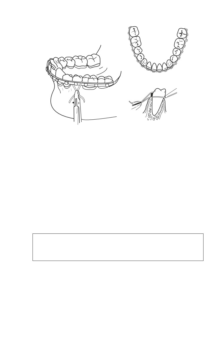
Place commercially made arch bars onto the facial aspect
of the maxillary and mandibular teeth.
♦ The arch-bars are then fixed to the teeth with simple
circumdental (24 or 25 gauge) wires (Fig. 13-1).
♦ After proper occlusion is established, the maxillary arch
bar is fixed to the mandibular arch bar with either wire
or elastics.
♦ If the patient’s jaws are wired together, it is imperative
that wire cutters be with the patient at all times.
♦ If portions of the mandible have been avulsed or the mandib-
ular fragments are extremely contaminated, an external
biphase splint should be placed to maintain alignment.
13.4
Face and Neck Injuries
Fig. 13-1. Arch bar applications.
ο Open reduction and internal fixation with a mandibular
plate across fracture sites may obviate the need for MMF.
Nasal fractures.
ο Most common fracture.
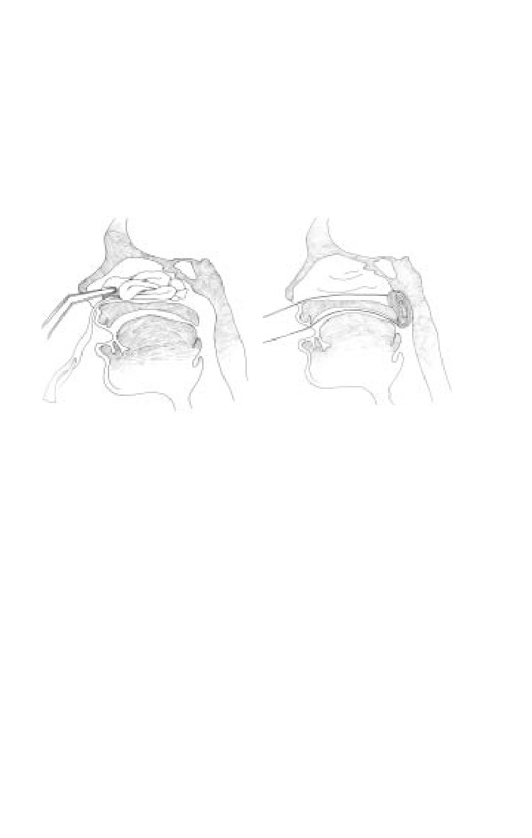
♦ Control of epistaxis: anterior pack-gauze/balloon/
tamponade.
ο Diagnosed clinically by the appearance and mobility of
the nasal bones.
The patient’s septum should be evaluated for the presence
of a septal hematoma, which if present, must be
immediately drained by incision, followed by packing.
ο Treat by closed reduction of the fractured bones and/or
septum into their correct anatomic positions up to 7 days
after fracture.
13.5
Emergency War Surgery
♦ Place a blunt elevator (Sayer) into the nasal cavity in
order to elevate the depressed bony segment while
simultaneously repositioning the bone with the
surgeon’s thumb placed externally.
ο The nose may then be fixed with tape or a splint in order
to maintain the reduction (Fig. 13-2).
a
b
Fig. 13-2. (a) Anterior and (b) posterior packing of the nose.
Maxillofacial Trauma.
ο Life-threatening due to loss of airway, hemorrhage, or
spinal injury.
ο Fragment wound of maxillary sinus is commonly seen and
requires surgical removal of retained fragments (can delay
until specialist is available).
Bleeding.
ο Common from epistaxis, oral hemorrhage or combination
bleeding.
ο Nasal fracture—most common fracture.
♦ Control Epistaxis with Anterior Pack (gauze/balloon/
tampon).
ο Mandibular fracture fixation (wires/archbars, with wire
cutters at bedside).
ο Facial and scalp lacerations.
ο Mid-face fracture (Le Fort)—The most difficult bleeding
to control.
13.6
Face and Neck Injuries
♦ Requires “significant” trauma.
♦ Be aware of associated CNS and orbital injury.
♦ Significant hemorrhage due to laceration of IMA and
branches.
◊ Is difficult to control.
◊ May be life-threatening.
◊ Treat by controlling airway, reducing fracture, and
placing a pressure dressing such as packing or
balloon.
♦ Edema may cause loss of airway, which may be
immediate or delayed.
♦ Can be difficult to diagnose. Criteria:
◊ Mobile hard palate and mid-face while stabilizing the skull.
◊ Penetrating injury may not follow classic Le Fort
patterns but may have a significant soft tissue injury
component (base of tongue, soft palate).
Treatment.
ο ABCs.
ο Check CNS and vision.
ο Can immobilize maxilla by using the mandible as a splint
(wires/archbars, with wire cutters at bedside).
ο Control hemorrhage by tamponade.
♦ Nasopharynx, nasal cavity.
♦ Oropharynx.
Surgical Repair.
ο Not an emergency once hemorrhage is controlled.
ο Requires ENT, oral, plastic, and ophthalmology surgical
expertise.
ο Time consuming.
ο Open and closed reductions with hardware that is usually
unavailable in the field.
Fractures of Facial Bones.
ο Potentially life-threatening due to loss of airway,
hemorrhage, or spinal injury.
ο Fragment wound of maxillary sinus is commonly seen,
and requires surgical removal of retained fragments (can
delay until specialist available).
ο Mid-face fracture (Le Fort)—The most difficult bleeding to
control.
♦ Requires “significant” trauma.
13.7

Emergency War Surgery
♦ Be aware of associated CNS and orbital injury.
♦ Significant hemorrhage due to laceration of IMA and
branches.
◊ Is difficult to control.
◊ May be life threatening.
◊ Treat by controlling the airway, reducing fractures,
and placing pressure dressings such as packing or
balloon tamponade.
♦ Edema may cause loss of airway, which may be
immediate or delayed.
♦ Can be difficult to diagnose.
◊ Mobilize the hard palate and mid-face while stabilizing
the skull. Place thumb and forefinger of one hand on
nasal bridge to stabilize, then with the other hand,
determine mobility of maxilla by placing the thumb on
alveolus and forefinger on the palate and attempting
gentle distraction in an anterior-posterior direction.
◊ Penetrating injury may not follow classic Le Fort
patterns but may have a significant soft tissue injury
component (base of tongue, soft palate).
◊ Apply principles of systemic palpation and
inspection, looking for crepitus, tenderness, internal
and external ecchymosis, and subconjunctival
hemorrhage that might suggest fractures.
ο
Classification by Le Fort (Fig. 13-3).
a
b
c
I
II
III
Fig. 13-3. Le Fort facial fracture classifications.
13.8
Face and Neck Injuries
♦ I-Fracture separates the entire alveolar process from
maxilla.
♦ II-Separation of mid-face, including the nasal bone,
from the orbit (pyramidal).
♦ III-Detachment of the face from the skull (craniofacial
disarticulation).
ο
Treatment.
♦ ABCs.
♦ If nasal intubation is used, extremely careful placement
is mandatory to avoid cribriform plate or anterior
cranial fossa penetration.
♦ Classification by Le Fort.
♦ Check CNS and vision.
♦ Can immobilize maxilla by using the mandible as a splint
(wires/arch bars, with wire cutters at bedside). It is much
easier to place patient into maxillomandibular fixation if
either a nasal airway or tracheostomy is employed.
♦ Control hemorrhage by tamponade as previously
described.
◊ Nasopharynx, nasal cavity.
◊ Oropharynx.
ο
Definitive Surgical Repair.
♦ Not an emergency once hemorrhage is controlled.
♦ Requires expertise in ENT, oral and maxillofacial,
plastic, and ophthalmology surgery.
♦ Time consuming.
♦ Open and closed reductions require hardware usually
unavailable in the field.
Soft Tissue Injuries
General principles.
ο Avoid injury to surrounding structures such as the facial
nerve or parotid duct.
ο Wounds should be gently cleansed with saline and light
scrub solutions; foreign bodies should be meticulously
cleaned from wounds prior to closure.
ο Sharply debride devascularized wound edges minimally.
ο Facial lacerations should be closed in layers within 24 hours.
♦ Use 4-0 or 5-0 absorbable suture for subcutaneous/
dermal layers.
13.9
Emergency War Surgery
♦ Use 5-0 or 6-0 nonabsorbable sutures on the skin of the face.
♦ Remove sutures in 5-7 days.
Facial nerve injuries.
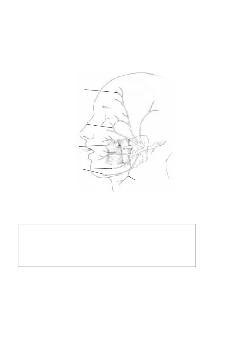
ο Carefully examine for facial nerve function in all five
branches (Fig. 13-4).
Temporal Branches
Zygomatic Branches
Buccal Branches
Marginal
Mandibular Branch
Cervical Branch
Fig. 13-4. Branches of the facial nerve parotid duct injury.
Facial nerve branches that are lacerated at a site anterior to
a vertical line drawn down from the lateral canthus of the
eye do not need to be surgically reapproximated because
these branches are very small and will spontaneously
regenerate with good return of facial function.
ο The severed ends of the nerve may be located in the wound
with a nerve stimulator, for up to 3 days.
ο Cut nerve ends should be reapproximated primarily with three
or four fine (9-0) nylon sutures placed through the epineurium.
ο If a gap exists between severed ends of the facial nerve due
to tissue loss, an interposition graft may be placed using a
section of the great auricular nerve to bridge the gap.
13.10
Face and Neck Injuries
ο If the wound is heavily contaminated and cannot be closed
primarily, the severed ends of the nerve should be located
and tagged for identification and repair at the time of
wound closure.
Parotid duct injuries.
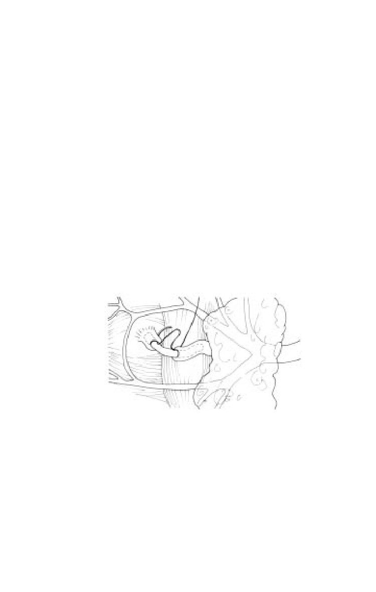
ο Evaluate penetrating wounds of the parotid/buccal
regions of the face for salivary leakage due to a lacerated
parotid duct (see Fig 13-5).
♦ The wound may be manually compressed and inspected
for salivary leakage.
♦ If the parotid duct is injured by a facial laceration, the
distal end of the duct may be identified by placing a
lacrimal probe through the intraoral opening of the duct
located near the maxillary second molar (see Fig. 13-4).
♦ The proximal end may be identified by compressing
the wound and identifying any areas of salivary
leakage.
ο Repair with absorbable (6-0) sutures (Fig. 13-5).
Fig. 13-5. Repair of parotid duct.
ο A stent may be placed into the duct to facilitate the closure
and prevent stenosis.
♦ Possible stents include lacrimal stents, large (size 0)
polypropylene sutures, or long angiocaths.
♦ Stents may be sutured to the buccal mucosa and
removed after seven days.
13.11
Emergency War Surgery
Penetrating Neck Trauma
Introduction.
ο Vascular injuries occur in 20% and aerodigestive tract in
10% of cases.
ο Mortality is primarily due to exsanguinating hemorrhage.
ο Esophageal injury, which results in mediastinitis and
intractable sepsis, may also be fatal.
Anatomy.
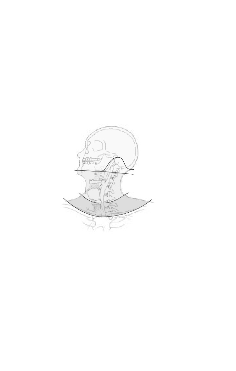
The neck is divided into three zones to aid decision making for
diagnostic tests and surgical strategy. In each zone, the primary
structures at risk of injury are different (Fig. 13-6).
Zone 3
Zone 2
Zone 1
Fig. 13-6. Zones of the neck.
ο Zone 1 (clavicle to cricoid membrane): The structures of
concern include large vessels of the thoracic outlet
(subclavian artery and vein, common carotid artery), the
lung, and the brachial plexus.
ο Zone 2 (cricoid membrane to angle of mandible): Structures
of concern include the common carotid artery, internal
jugular vein, esophagus, and trachea.
ο Zone 3 (angle of mandible to base of skull): The structure
of concern is primarily the internal carotid artery.
13.12
Face and Neck Injuries
Immediate management.
ο Initially, same as above.
ο Obtain chest and soft tissue neck radiographs.
ο Address tetanus and antibiotic prophylaxis.
Operative strategy.
ο If no platysma violation, surgical intervention is not
indicated.
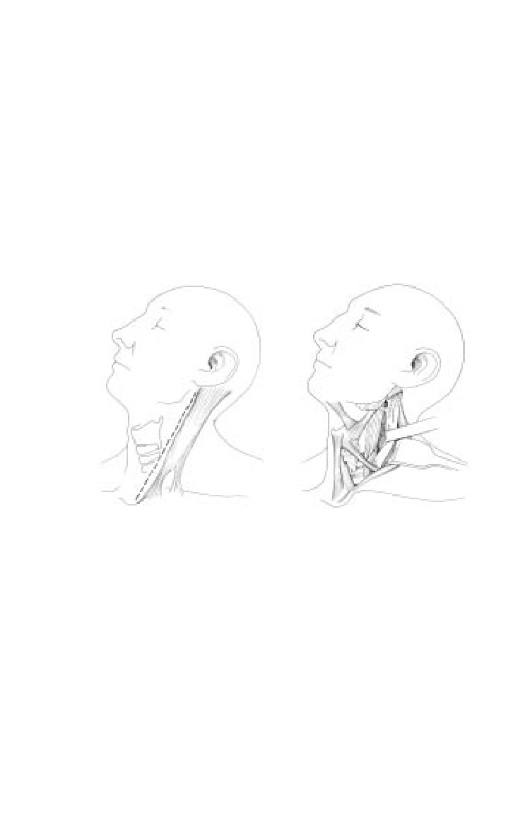
ο Zone 2 injuries that penetrate the platysma should undergo
routine exploration to rule out life threatening vascular,
esophageal or tracheal injuries via an incision along the anterior
border of the sternocleidomastoid muscle (Fig. 13-7).
Fig. 13-7. Neck exposure of zone 2.
ο Zone 1 and 3 injuries require selective management, based
on clinical signs and chest radiograph findings, making
an incision dependent on the vascular structure most
probably injured.
♦ Zone 1 and 3 penetrations without clinical signs of
injury (see below) may be evacuated without operative
intervention.
ο The most important clinical signs pointing to probable
injuries (pertinent to all 3 zones):
13.13
Emergency War Surgery
♦ Signs of vascular injury.
◊ Current or history of significant bleeding.
◊ Expanding hematoma.
◊ Bruit or thrill in the neck.
◊ Hypotension.
◊ Dyspnea, hoarseness, or stridor.
◊ Absent or decreased pulses in neck or arm.
◊ Focal neurologic deficit or mental status change.
◊ Chest radiograph findings of hemothorax or
mediastinal widening.
♦ Signs of aerodigestive injury (esophagus, trachea,
larynx).
◊ Crepitus or subcutaneous emphysema.
◊ Dyspnea or stridor.
◊ Air bubbling from wound.
◊ Tenderness or pain over trachea; odynophagia.
◊ Hoarse or abnormal voice.
◊ Hematemesis or hemoptysis.
Surgical Principles
The groin and upper thigh should be surgically prepped for
greater saphenous vein interposition graft or patch angioplasty.
Exsanguinating hemorrhage from injured vessels at the base
of the skull (Zone 3) can often be controlled with inflation of
a directed catheter (Fogarty, Foley), left in place and inflated
for 48-72 hours, then deflated in the OR under controlled
visualization for rebleeding.
Repair esophageal injuries in a single-layer and place closed
suction drains. The drain tip should not be placed near a
concomitantly repaired carotid artery. A muscle flap should
be interposed between repaired esophageal and tracheal
injuries to prevent fistula. Obtain an oral contrast swallow
radiograph seven days after repair before feeding.
Repair laryngotracheal injuries with single-layer
monofilament absorbable suture. Must search for
concomitant esophageal injuries.
Unreconstructable (significant segmental loss, or > 50%
diameter loss) tracheal injuries should be managed with an
endotracheal tube placed through the defect.
13.14
Face and Neck Injuries
Vertebral artery injury.
ο Suspect if bleeding continues from a posterolateral neck
wound despite pressure on the carotid artery.
ο Preoperative angiography localizes site of injury and estab-
lishes the existence of a patent contralateral vertebral artery,
aplasia of which is most commonly located on the left side.
ο Exposure of vertebral artery may be difficult. When
contralateral vertebral artery is intact, ligation proximal
and distal to the injury will likely be necessary.
ο Bone wax or a Foley catheter may be useful for control of bleeding.
Intraoral injuries
ο Penetrating injuries to the oral cavity LATERAL to the
tonsillar fossa are at a significant risk of causing occult
internal carotid injury. Neurologic testing/monitoring is
critical and CT scanning and/or angiography should be
considered. If after a penetrating lateral oral injury the
patient bleeds a small amount only to stop, this may signify
a “sentinel” bleed. A carotid blowout may follow.
Internal carotid artery injury.
ο
Should be repaired primarily unless there is profound
hemiplegia with deep coma Glasgow Coma Scale (GCS <
8). All other carotid branches can be ligated.
ο
The use of carotid shunts during repair has no proven benefit.
ο
In small perforations, debride minimally, and close with
6-0 polypropylene.
ο
With loss of vascular tissue, vein angioplasty is required.
ο
If there is extensive destruction, segmental resection and
restitution of flow is established by:
♦ End-to-end anastomosis (if the vessel is sufficiently
elastic to permit).
♦ Interposition vein graft.
♦ External carotid swing-over and interposition.
♦ Temporary (24-48 h) shunt as part of damage control
maneuver.
ο
The mortality is high in patients with severe neurologic deficit;
carotid ligation is justifiable in complete occlusion of the entire
carotid system and depending on the triage situation.
ο
Distal clot may be removed by extremely gentle use of a
balloon catheter prior to shunt insertion or repair.
13.15
Emergency War Surgery
Internal jugular vein injury.
ο
Preferably repaired by lateral suture.
ο
Ligation OK, if the contralateral internal jugular is patent.
ο
Larynx.
ο
After immediate control of the airway has been achieved
by intubation or tracheotomy (not through the wound in
the larynx!), a complete airway evaluation by direct
laryngoscopy and bronchoscopy must be performed.
ο
Debridement of laryngotracheal injuries must be careful
and conservative. A fragmented larynx or trachea should
be reapproximated and sutured with extraluminal,
absorbable sutures for tracheal injuries and nonabsorbable
sutures or micro-plates used for laryngeal fractures.
ο
The management of laryngeal trauma includes accurate
reduction and stabilization of fractures, mucosa-to-mucosa
closure of lacerations, and use of a soft stent if there is
extensive cartilaginous damage and structural support is
decreased or the anterior commissure is involved. The stent
may need to be temporarily placed for 4-6 weeks to
maintain correct anatomic architecture and requires a
complementary tracheotomy.
ο
The excessive removal of cartilage and mucosa must be
avoided to prevent tracheal or laryngeal stenosis.
Laryngotracheal injuries.
ο If laryngotracheal separation is suspected (massive crepitis
over the larynx/trachea) in an otherwise “stable” airway,
endotracheal intubation should not be undertaken as this
may cause a partial separation to become a complete
separation and/or blind passage of the endotracheal tube
may occur with resulting impending airway emergency.
ο It is best to perform an awake tracheotomy/
cricothyroidotomy under local anesthesia without
paralysis. Good anesthesia can be achieved with a 4%
(40mg/cc) lidocaine nebulizer, 2cc in 3cc of saline, and
direct administration of 4% lidocaine into the trachea for
an awake tracheotomy (in addition to local anesthetic
infiltration into the skin and subcutaneous tissues). When
instilling anesthesia into the airway, aspirate and ensure
air enters the syringe before injecting.
13.16
Face and Neck Injuries
Tracheal injury and reconstruction.
ο Small anterior wounds can have tracheostomy tube placed
through them after debridement.
ο Repair simple lacerations with absorbable monofilament
suture.
ο Up to 5 cm can be resected with proximal and distal
mobilization.
ο Mobilize anteriorly and posteriorly to preserve lateral
blood supply.
ο Remove endotracheal tube as soon as possible post-op.
ο May need to suture chin to chest for 10 days to avoid
extension injury.
Esophageal injury and repair.
ο Difficult to diagnose.
ο 25% may be asymptomatic.
ο Missed injury is a major source of late morbidity/mortality.
ο Insufflation with air may aid in identification during
exploration.
ο Debride devitalized tissue.
ο Wound closure in two layers with absorbable sutures.
ο Viable muscle flap to protect repairs from leak.
ο Drainage with closed suction drain.
ο Barium swallow 7 days post-op, prior to oral intake.
ο Oral intake prior to drain removal.
ο Extensive injuries may require lateral cervical
esophagostomy.
ο Cervical esophagostomy is preferred to closure under
tension.
Combined injuries.
ο All esophageal injuries combined with airway or vascular
injury require separation with healthy tissue. Strap muscles
are ideal, but can use pedicle of sternocleidomastoid if
straps are devitalized.
Esophageal fistula.
ο 10%-30% incidence.
ο Due to inadequate debridement, devascularization of
remaining esophageal wall, closure under tension, or
infection.
ο Treatment.
13.17
Emergency War Surgery
♦ Maintain nutrition.
♦ Assure control with drains.
♦ Weekly barium swallow to assess closure.
♦ Oral intake prior to removing drain.
Skull Base, Temporal Bone and Otologic Injury
Ensure that the facial nerve is assessed and documented on
an awake patient and at the earliest convenience in a patient
who has regained consciousness. Delineation between
delayed onset and acute facial paralysis is critical for
management and outcome of facial nerve injuries. Also critical
is delineation between a distal and proximal nerve injury. If
a distal injury is present, one or more branches may be
affected.
ο Be as concise as possible in describing facial motion even
if not technically accurate; be complete in the description.
A more proximal injury (proximal to the Pes Anserinus)
will most likely result in all branches being equally
affected. Accurate documentation may spare the patient
from unwarranted surgical intervention to explore the
entire length of the facial nerve. It is desirable to accurately
describe the motion of EACH branch of the facial nerve.
Eyelid movement does not ensure that the facial nerve is
intact since the levator palpebrae is innervated by the
occulomotor nerve and will remain intact despite facial
nerve injury.
ο If there is no contraindication for systemic steroids, they
should be administered for suspected facial nerve
paralysis. Crush injuries to the facial nerve may present
with delayed onset paralysis and the severity and course
of the paresis may be improved with systemic
administration of steroids.
Skull base fractures are often occult. Assess the patient for
evidence of basilar skull fractures (Battle’s sign, raccoon eyes).
Ensure that the external auditory canal is examined. Do not
instrument the external auditory canal, however. If a temporal
bone fracture is present and the dura is not intact,
instrumentation may introduce bacteria and/or a foreign
body into the CSF.
13.18
Face and Neck Injuries
The external auditory canal should be inspected for a tear of
the lining of the canal. A tear of the lining of the canal suggests
a temporal bone fracture.
ο If a temporal bone fracture is suspected, it is critical that
the facial nerve be assessed.
Tympanic membrane perforations can be managed
expectantly. The vast majority of them will heal
spontaneously, but the patient should be followed for
evidence of cholesteatoma formation from traumatic
implantation of the squamous epithelium. This may occur
months to years after the injury. Acutely, application of otic
antibiotic drops will prevent the perforation from desiccating,
but this is not required. The patient should be instructed to
keep the ears dry (avoid water contamination).
Hemotympanum may be seen with acoustic & temporal bone
trauma. These patients will have hearing loss. If available,
perform a gross audiological evaluation with tuning forks.
Hemotympanum with hearing loss (conductive) should
resolve in about 6 weeks.
ο Examination for hearing in the field can be accomplished
with a single 512 tuning fork.
♦ With the tuning fork placed on the mastoid tip and then
alternately in front of the external canal (Rinne).
Documentation as A>B (air > bone) or B>A is sufficient
- do not report as “positive” or “negative”:
◊ Air conduction greater than bone conduction with a
512 tuning fork is normal.
◊ Bone conduction greater than air is suggestive of a
conductive hearing loss.
♦ With the 512 tuning fork on the frontal bone/ nasal
dorsum/ or central incisors (best) (Weber):
◊ If the Rinne test suggests a conductive hearing loss -
the 512 should lateralize to the side with the
conductive loss.
◊ If the Rinne is NORMAL (A>B) - the 512 should
lateralize to the ear with a sensorineural loss
Any otologic blast injury or injury to the temporal bone may
result in tinnitus. Management is expectant and it may resolve
13.19
Emergency War Surgery
spontaneously. Accurate documentation is critical for future
management of these patients, however.
If sensorineural hearing loss is suspected and documented
after a blast injury or noise trauma, steroids are indicated.
1mg/kg of prednisone is appropriate. If after five days there
is no improvement, the patient can be taken off of the steroids.
If improvement is noted, a taper over 3-4 weeks is indicated.
Be mindful that steroids may affect a patient’s affect and
impair judgment.
Dizziness and vertigo may result from acoustic trauma. If
true vertigo exists after an otologic injury (observed
nystagmus), the patient may have a perilymphatic fistula
from depression of the stapes into the oval window or
rupture of the round window. These patients may have
tinnitus and hearing loss with vertigo. If a perilymphatic
fistula is suspected, this patient should be seen by an
Otolaryngologist as soon as possible to prevent further
damage to the inner ear.
13.20
Chapter 14
Ocular Injuries
Introduction
The preservation of the eyes and eyesight of service personnel
is an extremely important goal. Despite comprising as little as
0.1% of the total body surface area, injuries to the eye are found
in 5-10% of all combat casualties. In the Vietnam War almost
50% of casualties with penetrating eye wounds eventually lost
vision in the injured eye. Improvements in ophthalmic care in
the last 30 years offer hope that blindness in combat casualties
will be less common in future wars.
Triage of Patients With Eye Injuries
ABCs (airway, breathing, and circulation) and life-threatening
injuries have priority, then treat eyesight and limbs.
Soldiers with mild eye injuries may be treated and returned
to duty by nonspecialized personnel.
Soldiers with more severe injuries should be evacuated to
save vision.
Distinguishing major ocular injuries from minor ones may
be difficult.
At the FST level, due to time and equipment restraints,
surgeons will likely ‘patch and evacuate’.
Identifying Severe Eye Injuries
Associated injuries.
ο Shrapnel wounds of the face — think intraocular foreign
body (IOFB).
ο Lid laceration — check for underlying globe laceration.
Vision.
14.1
Emergency War Surgery
ο Use book print, medication labels, finger counting, and
the like, to evaluate vision.
ο Compare sight in the injured eye to the uninjured eye.
ο Severe vision loss is a strong indicator of serious injury.
Eyeball structure.
ο Obvious corneal or scleral lacerations.
ο Subconjunctival hemorrhage — may overlay an open
globe.
ο Dark uveal tissue presenting on the surface of the eye
indicates an open globe.
ο Foreign body — did it penetrate the eye?
ο Blood in the anterior chamber (hyphema) indicates severe
blunt trauma or penetrating trauma.
Proptosis — may indicate a retrobulbar hemorrhage.
Pupils.
ο Pupillary distortion — may be associated with an open
globe.
Motility.
ο Decreased motility on one side may be caused by an open
globe.
ο Other causes include muscle injury, orbital fracture, and
orbital hemorrhage.
Open Globe
May result from penetrating or blunt eye trauma.
May cause loss of vision from either disruption of ocular
structures or secondary infection (endophthalmitis).
Biplanar radiographs or a CT (computed tomagraphy) scan
of the head may help to identify a metallic intraocular
fragment in a casualty with severe vision loss, a traumatic
hyphema, a large subconjunctival hemorrhage, or other signs
suspicious for an open globe with an IOFB.
Immediate Treatment of an Open Globe
Tape a rigid eye shield (NOT a pressure patch) over the eye.
Do not apply pressure on or manipulate the eye.
Do not apply any topical medications.
Start quinolone antibiotic PO or IV (eg, ciprofloxacin 500 mg bid).
14.2
Ocular Injuries
Schedule an urgent (within 24-48 h) referral to an ophthal-
mologist.
Administer tetanus toxoid if indicated.
Prevent emesis (Phenergan 50 mg or Compazine 10 mg IM/IV).
Treatment of Other Anterior Segment Injuries
Subconjunctival Hemorrhage
Small subconjunctival hemorrhages (SCH) may occur
spontaneously or in association with blunt trauma. These
lesions require no treatment.
SCH may also occur in association with a rupture of the
underlying sclera.
Warning signs for an open globe include a large SCH with
chemosis (conjunctiva bulging away from globe) in the setting
of blunt trauma, or any SCH in the setting of penetrating
injury. Casualties with blast injury and normal vision do not
require special care.
Suspected open globe patients should be treated as described
above.
Treatment of Chemical Injuries of the Cornea
Immediate copious irrigation (for 30 minutes) with normal
saline (NS), lactated Ringer’s (LR), or balanced salt solution.
Nonsterile water may be used if it is the only liquid available.
Use topical anesthesia before irrigating, if available.
Measure the pH of tears to ensure that if there is either acid
or alkali in the eye, the irrigation continues until the pH
returns to normal. Do not use alkaline solutions to neutralize
acidity or vice versa.
Remove any retained particles.
Using fluorescein test, look for epithelial defect.
ο If none, then mild chemical injuries or foreign bodies may
be treated with artificial tears.
ο If an epithelial defect is present, use a broad-spectrum
antibiotic ophthalmic ointment (Polysporin, erythromycin,
or bacitracin) 4 times per day.
Noncaustic chemical injuries usually resolve without
sequelae.
14.3
Emergency War Surgery
More severe chemical injuries may also require treatment with
prednisolone 1% drops 4-9 times per day and scopolamine
0.25% drops 2-4 times per day.
Pressure patch between drops or ointment if a large epithelial
defect is present.
Monitor (daily topical fluorescein evaluation) for a corneal
ulcer until epithelial healing is complete.
Severe acid or alkali injuries of the eye (recognized by
pronounced chemosis, limbal blanching, and/or corneal
opacification) can lead to infection of the cornea, glaucoma,
and possible loss of the eye. Refer to an ophthalmologist
within 24-48 hours.
Treat mustard eye injuries with ophthalmic ointments, such
as 5% boric acid ointment, to provide lubrication and minimal
antibacterial effects. Apply sterile petrolatum jelly between
the eyelids to provide additional lubrication and prevent
sealing of the eyelids.
Treat nerve agent ocular symptoms with 1% atropine sulfate
ophthalmic ointment, repeat as needed at intervals of several
hours for 1-3 days.
Corneal Abrasions
Diagnosis.
ο Be alert for the possibility of an associated open globe.
ο The eye is usually very symptomatic with pain, tearing,
and photophobia.
ο Vision may be diminished from the abrasion itself or from
the profuse tearing.
ο Diagnose with topical fluorescein and cobalt blue light
(Wood’s lamp).
ο A topical anesthetic may be used for diagnosis, but should
NOT be used as an ongoing analgesic agent — this delays
healing and may cause other complications.
Treatment.
ο Apply broad spectrum antibiotic ointment (Polysporin,
erythromycin, or bacitracin) qid.
ο Options for pain relief.
♦ Pressure patch (usually sufficient for most abrasions).
♦ Diclofenac 0.1% drops qid.
14.4
Ocular Injuries
♦ Larger abrasions may require a mild cycloplegic agent
(1% Mydriacyl or Cyclogyl) and a pressure patch.
♦ More severe discomfort can be treated with 0.25%
scopolamine one drop bid, but this will result in pupil
dilation and blurred vision for 5-6 days.
ο Small abrasions usually heal well without patching.
ο If the eye is not patched
♦ Antibiotic drops (fluoroquinolone or aminoglycoside)
may be used qid in lieu of ointment.
♦ Sunglasses are helpful in reducing photophobia.
ο Casualties who wear contact lenses should have the lens
removed and should not be treated with a patch because
of the higher risk of developing a bacterial corneal ulcer.
ο Abrasions will normally heal in 1-4 days.
ο Initial treatment of thermal burns of the cornea is similar
to that for corneal abrasions.
All corneal abrasions need to be checked once a day until
healing is complete to ensure that the abrasion has not
been complicated by secondary infection (corneal ulcer,
bacterial keratitis).
Corneal Ulcer and Bacterial Keratitis
Diagnosis.
ο Corneal ulcer and bacteria keratitis are serious conditions
that may cause loss of vision or even loss of the eye!
ο A history of corneal abrasion or contact lens wear.
ο Increasing pain and redness.
ο Decreasing vision.
ο Persistent or increasing epithelial defect (positive
fluorescein test).
ο White or gray spot on the cornea seen on examination with
penlight or direct ophthalmoscope.
Treatment.
ο Quinolone drops (eg, Ocuflox), 1 drop every 5 minutes
for 5 doses initially, then 1 drop every 30 minutes for 6
hours, then 1 drop hourly around the clock thereafter.
ο Scopolamine 0.25%, one drop bid may help relieve
discomfort caused by ciliary spasm.
14.5
Emergency War Surgery
ο Patching and use of topical anesthetics for pain control
are contraindicated (see pain control measures above).
ο Expedited referral to an ophthalmologist within 3-5 days
unless patient is not improving within 48 hours. Infection
may worsen, leading to permanent injury.
Conjunctival and Corneal Foreign Bodies
Diagnosis.
ο Abrupt onset of discomfort and/or history of suspected
foreign body.
ο If an open globe is suspect, treat as discussed above.
ο Definitive diagnosis requires visualization of the offending
object, which may sometimes be quite difficult.
♦ A hand-held magnifying lens or pair of reading glasses
will provide magnification to aid in the visualization
of the foreign body.
♦ Stain the eye with fluorescein to check for a corneal
abrasion.
ο The casualty may be able to help with localization if asked
to indicate the perceived location of the foreign body prior
to instillation of topical anesthesia.
ο Eyelid eversion with a cotton-tipped applicator helps the
examiner identify foreign bodies located on the upper
tarsal plate.
Treatment.
ο Superficial conjunctival or corneal foreign bodies may be
irrigated away or removed with a moistened sterile swab
under topical anesthesia.
ο Objects adherent to the cornea may be removed with a
spud or a sterile 22-gauge hypodermic needle mounted
on a tuberculin syringe (hold the needle tangential to the
eye).
ο If no foreign body is visualized, but the index of suspicion
is high, vigorous irrigation with artificial tears or sweeps
of the conjunctival fornices with a moistened cotton-tipped
applicator after topical anesthesia may be successful in
removing the foreign body.
ο If an epithelial defect is present after removal of the foreign
body, treat as discussed above for a corneal abrasion.
14.6
Ocular Injuries
Hyphema: Blood in the Anterior Chamber
Treatment (to prevent vision loss from increased intraocular
pressure).
ο
Be alert for a possible open globe and treat for that
condition if suspected.
ο
Avoidance of rebleeds is a major goal of management.
♦ Avoid aspirin or nonsteroidal antiinflammatory drugs
(NSAID).
♦ No strenuous activity (bedrest) for 14 days.
♦ No reading for 7 days.
ο
Prednisolone 1% drops 4 times a day.
ο
Scopolamine 0.25% drops twice a day.
ο
Cover eye with protective shield.
ο
Elevate head of bed to promote settling of red blood cells
(RBC) in anterior chamber.
ο
Provide a 24-48 hour referral to an ophthalmologist to
monitor for increased intraocular pressure (which may
cause permanent injury to the optic nerve) and to evaluate
for an associated open globe.
ο
If evaluation by an ophthalmologist is delayed (> 24 hrs),
treat with a topical B-blocker (timolol or levobunolol) bid
to help prevent intraocular pressure elevation.
ο
If intraocular pressure is found to be markedly elevated
(above 30 mm Hg) with a tonopen or other portable
tonometry device, other options for lowering intraocular
pressure include acetazolamide 500 mg PO or IV and
mannitol 1-2 g/kg IV over 45 minutes.
Retrobulbar (Orbital) Hemorrhage
Keys to recognition: Severe eye pain, proptosis, vision loss,
decreased eye movement.
ο Marked lid edema may make the proptosis difficult to
appreciate.
ο Failure to recognize may result in blindness from increased
ocular pressure.
Perform an immediate lateral canthotomy.
Provide an urgent referral to an ophthalmologist, within 24-
48 hours.
14.7
Emergency War Surgery
If evaluation by an ophthalmologist is delayed (>24 hrs), treat
with a topical B-blocker (timolol) bid to help lower intraocular
pressure elevation.
If intraocular pressure is found to be elevated (>30 mm Hg),
treat as discussed above.
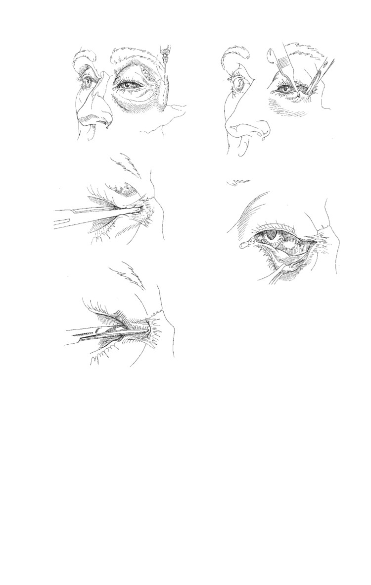
Lateral Canthotomy/Cantholysis
Do not perform such procedures if the eyeball structure has been
violated. If the eye is sliced open, apply a Fox shield for
protection and seek immediate ophthalmic surgical support.
Inject 2% lidocaine with 1:100,000 epinephrine into the lateral
canthus (Fig. 14-1a).
Crush the lateral canthus with a straight hemostat, advancing
the jaws to the lateral fornix (Fig. 14-1b).
Using straight scissors make a 1-cm long horizontal incision
of the later canthal tendon, in the middle of the crush mark
(Fig. 14-1c).
Grasp the lower eyelid with large toothed forceps pulling
the eyelid away from the face. This pulls the inferior crus
(band of the lateral canthal tendon) tight so it can be easily
cut loose from the orbital rim (Fig.14-1d).
ο
Use blunt tipped scissors to cut the inferior crus.
ο
Keep the scissors parallel (flat) to the face with the tips
pointing toward the chin.
ο
Place the inner blade just anterior to the conjunctiva and
the outer blade just deep to the skin.
ο
The eyelid should pull freely away from the face, releasing
pressure on the globe.
ο
Cut residual lateral attachments of the lower eyelid if it
does not move freely.
ο
Do not worry about cutting 1/2 cm of conjunctiva or skin.
ο
The lower eyelid is cut, relieving orbital pressure. If the
intact cornea is exposed, apply, hourly, copious
erythromycin ophthalmic ointment or ophthalmic
lubricant ointment to prevent devastating corneal
dessication and infection. Relief of orbital pressure must
be followed by lubricating protection of the cornea and
urgent ophthalmic surgical support. Do NOT apply
absorbent gauze dressings to the exposed cornea.
14.8
Ocular Injuries
a
d
b
e
c
Fig. 14-1. Lateral canthotomy and inferior cantholysis are indicated
for casualties presenting with serious orbital hemorrhage.
Orbital Floor (Blowout) Fractures
These fractures are usually the result of a blunt injury to the
globe or orbital rim, often associated with head and spine
injuries. Blowout fractures may be suspected on the basis of
enophthalmos, diplopia, decreased ocular motility, hypoesthesia
of the V2 branch of the trigeminal nerve, associated subcon-
junctival hemorrhage, or hyphema. Immediate treatment
includes pseudoephedrine 60 mg q 6 hours and a broad-
14.9
Emergency War Surgery
spectrum antibiotic for 7 days, ice packs, and instructing the
casualty not to blow his nose. Definitive diagnosis requires CT
scan of orbits with axial and coronal views. Indications for repair
include severe enophthalmos and diplopia in the primary or
reading gaze positions. The surgery may be performed 1-2
weeks after the injury.
Lid Lacerations
Treatment guidelines for lid lacerations not involving the
lid margin
Excellent blood supply — delayed primary closure is not
necessary.
Eyelid function (protecting the globe) is the primary
consideration.
Begin with irrigation, antisepsis (any topical solution), and a
check for retained foreign bodies.
Superficial lacerations of the eyelid, not involving the eyelid
margin, may be closed with running or interrupted 6-0 silk
(preferred) or nylon sutures.
Horizontal lacerations should include the orbicular muscle
and skin in the repair.
If skin is missing, an advancement flap may be created to fill
in the defect. For vertical or stellate lacerations, use traction
sutures in the eyelid margin for 7-10 days.
Antibiotic ointments qid.
Skin sutures may be removed in 5 days.
Treatment guidelines for lid lacerations involving the lid margin
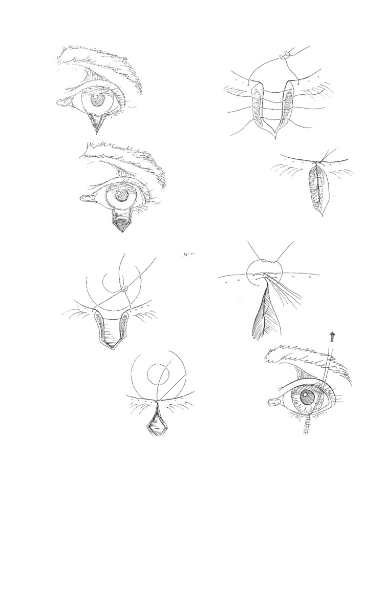
Repair of a marginal lower-eyelid laceration with less than
25% tissue loss (Fig. 14-2).
ο The irregular laceration edges may be freshened by
creating a pentagonal wedge — remove as little tissue as
possible (Fig. 14-2b).
ο A 4-0 silk or nylon suture is placed in the eyelid margin
(through the meibomian gland orifices 2 mm from the
wound edges and 2 mm deep) and is tied in a slipknot.
Symmetric suture placement is critical to obtain post-op
eyelid margin alignment (Fig. 14-2c).
14.10
Ocular Injuries
e
a
f
b
g
c
h
d
Fig. 14-2. Lid margin repair.
ο The slipknot is loosened and approximately two or three
absorbable (Vicryl or gut) 5-0 or 6-0 sutures are placed
internally to approximate the tarsal plate. The skin and
conjunctiva should not be included in this internal closure
(Fig. 14-2d).
14.11
Emergency War Surgery
ο Anterior and posterior marginal sutures (6-0 silk or nylon)
are placed in the eyelid margin just in front and behind
the previously placed 4-0 suture (Fig. 14-2e).
ο The middle and posterior sutures are left long and tied
under the anterior suture. Ensure that the wound edges
are everted (Fig. 14-2f).
ο The skin is closed with 6-0 silk or nylon sutures. The lid is
placed on traction for at least 5 days. The skin sutures are
removed at 3-5 days and the marginal sutures are removed
at 10-14 days (Fig. 14-2g).
Additional Points in Lid Laceration Repair
Tissue loss greater than 25% will require a flap or graft.
If there is orbital fat in the wound or if ptosis is noted in an
upper lid laceration, damage to the orbital septum and the
levator aponeurosis should be suspected.
If the eyelid is avulsed, the missing tissue should be retrieved,
wrapped in moistened Telfa, and preserved on ice. The tissue
should be soaked in a dilute antibiotic solution prior to
reattachment. If necrosis is present, minimal debridement
should occur in order to prevent further tissue loss. The
avulsed tissue should be secured in the anatomically correct
position in the manner described for lid margin repair above.
ο Damage to the canalicular system can occur as a result of
injuries to the medial aspect of the lid margins. Suspected
canalicular injuries should be repaired by an ophthal-
mologist to prevent subsequent problems with tear
drainage. This repair can be delayed for up to 24 hours.
Laser Eye Injuries
Battlefield lasers may be designed to cause eye injuries or
may be part of other weapons or sensor systems.
Prevention is the best option! Wear eye protection designed
for the appropriate light wavelengths if there is a known laser
threat.
The type of ocular damage depends on the wavelength of
the laser — retinal injuries are most common.
14.12
Ocular Injuries
The primary symptom of laser injury is loss of vision, which
may be preceded by seeing a flash of light. Pain may not be
present.
Immediate treatment of corneal laser burns is similar to that
for corneal abrasions.
Laser retinal burns have no proven immediate treatment,
although improvement with corticosteroids has been
reported.
Routine evacuation for evaluation by an ophthalmologist is
required.
Enucleation
A general surgeon in a forward unit should not remove a
traumatized eye unless the globe is completely disorganized.
Enucleation should only be considered if the patient has a very
severe injury, no light perception using the brightest light source
available, and is not able to be evacuated to a facility with an
ophthalmologist. Sympathetic ophthalmia is a condition that
may result in loss of vision in the fellow eye if a severely
traumatized, nonseeing eye is not removed, but it rarely
develops prior to 21 days after an injury. Delaying the
enucleation until the patient can see an ophthalmologist is
thus relatively safe.
14.13
Chapter 15
Head Injuries
Introduction
The prognosis of brain injuries is good in patients who respond
to simple commands, are not deeply unconscious, and do not
deteriorate. The prognosis is grave in patients who are rendered
immediately comatose (particularly those sustaining penetrating
injury) and remain unconscious for a long period of time. Any
subsequent neurologic improvement may indicate
salvageability and should prompt reevaluation.
Neurosurgical damage control includes early intracranial
pressure (ICP) control; cerebral blood flow (CBF) preservation;
and prevention of secondary cerebral injury from hypoxia,
hypotension, and hyperthemia.
A motor examination of the most salvageable severely brain-
injured patients will demonstrate localization to central
stimulation and these patients will require expedited treatment.
Immediate intubation with adequate ventilation is the most
critical first line of treatment for a severely head-injured patient.
Evacuation to the nearest neurosurgeon, avoiding diagnostic
delays, and initiating cerebral resuscitation allow for the best
chance for ultimate functional recovery.
Combat Head Injury Types
Blunt (closed head injury).
Penetrating.
ο Penetrating with retained fragments.
ο Perforating.
ο Guttering (grooving the skull).
ο Tangential.
ο Cranial facial degloving (lateral temple, bifrontal).
15.1