Emergency War Surgery (2004) - page 4
Emergency War Surgery
Table 9-1. Induction Agents and Sedatives.
Agent
Routine Dose*
Characteristics
Concerns
Ketamine
1.0-2.0 mg/kg IV
Dissociative anesthetic
Varying degrees
and amnestic.
of purposeful
Sympathomimetic
skeletal move-
effects (useful in
ment despite
hypovolemia).
intense
Potent bronchodilator.
analgesia
and amnesia.
4.0-8.0 mg/kg
Onset within 30-60 sec.
Increased
IM
Emergence delirium
salivation,
avoided with con-
consider an
comitant benzodia-
antisialagogue.
zepine use.
Barbiturates
3-5 mg/kg
Onset within 30-60
May cause
(eg, thiopental)
seconds.
profound
hypotension in
hypovolemic
shock patients.
Propofol
1.5-2.5 mg/kg
Mixed in lipid, strict
Contraindicated
sterility must be
in acute
ensured.
hypovolemic
Rapid onset and rapidly
shock patients.
metabolized.
Onset within 30-60
seconds.
Etomidate
0.2-0.4 mg/kg
Onset within 30-60
May cause
seconds.
clonus.
Duration 3-10 min.
Minimal cardiac effects.
Minimal effects on
peripheral and
pulmonary circulation.
Maintains cerebral
perfusion.
* All induction agents can be used for induction of severely injured patients
if reduced dosages are used (eg, 1/2 of the lower recommended dose).
However, the recommended choice for hypovolemic patients would be
Ketamine > etomidate >> thiopental > propofol.
9.4
Anesthesia
Rapid Sequence Intubation (RSI) 7 steps*
1. Preoxygenate with 100% oxygen by mask.
2. Consider fentanyl—titrate to maintain adequate blood
pressure and effect (2.0-2.5 µg/kg).
3. Cricoid pressure—Sellick maneuver until endotracheal
tube (ETT) placement is confirmed and balloon is
inflated.
4. Induction agent: etomidate 0.1-0.4 mg/kg IV push.
5. Muscle relaxant: succinylcholine 1.0-1.5 mg/kg IV push.
6. Laryngoscopy and orotracheal intubation.
7. Verify tube placement.
*For children, see page 33.6.
Endotracheal intubation.
ο
Orotracheal.
♦ Direct laryngoscopy 60-90 seconds after administration
of induction agents and neuromuscular blockade.
♦ First attempt is the best chance for success, but have a
backup plan:
◊ Optimize positioning of patient and anesthesia
provider.
◊ Have adjuncts readily available (stylet, smaller
diameter tubes, alternative laryngoscope blades,
suction, laryngeal mask airway, lighted stylet).
ο
Nasotracheal should generally not be performed.
ο
Other considerations.
♦ Maintain cricoid pressure until balloon inflated and tube
position is confirmed.
♦ Hypertension can be managed with short-acting
medications such as beta blockers (labetalol, esmolol)
or sodium nitroprusside.
♦ May treat induction-related (transient) hypotension
initially with small dose of ephedrine (5-10mg) or
Neosynephrine (50 µg), but if hypotension persists after
induction agents are metabolized, use fluids to treat the
persistent hypovolemia. The anesthesiologist must
convey this situation to the surgeon, as the need to
control bleeding becomes urgent.
9.5
Emergency War Surgery
♦ A sensitive airway can be topically anesthetized with
lidocaine 1.5 mg/kg 1-2 minutes before laryngoscopy.
Verify ETT placement.
ο Auscultate the lungs.
ο Measure the end-tidal CO2.
ο Ensure that the SaO
remains high.
2
ο Palpate cuff of ETT in sternal notch.
ο Place the chemical CO2 sensors in the airway circuit.
Verification of tube placement is VITAL. Any difficulty
with oxygenation/ventilation following RSI should
prompt evaluation for immediate reintubation.
The Difficult Airway (see Chapter 5, Airway and Breathing)
Initially provide airway management with jaw-thrust, facemask
oxygenation, and assess the situation. Failed RSI may be due to
inadequate time for induction agents to work; inadequate time
for muscle relaxation to occur; anatomically difficult airway; or
obstruction due to secretions, blood, trauma, or foreign material.
Resume oxygenation; consider placing a temporary oral airway.
Reposition patient and anesthesia provider.
Call for help.
Consider alternatives to RSI.
ο Awake intubation.
ο Laryngeal mask airway.
ο Regional anesthesia or local anesthesia.
ο Surgical airway.
Maintenance of General Anesthesia
General Anesthesia Is Maintained After Intubation With
Oxygen. Titrate to maintain SaO
> 92%.
2
Ventilation.
ο Tidal volume (TV) 10-15 cc/kg.
ο Respiratory rate (RR) 6-10/min.
ο PEEP (positive end-expiratory pressure) if desired at 5 cm
H2O, titrate as necessary.
Minimal alveolar concentration (MAC).
ο 0.3-0.5 MAC: awareness abolished although 50% of
patients respond to verbal commands.
9.6
Anesthesia
ο 1 MAC: 50% of patients do not move to surgical stimulus.
ο 1.2 MAC: 95% of patients do not move to surgical stimulus.
ο Common inhalation agent MACs:
♦ Halothane: 0.75%.
♦ Sevoflurane: 1.8%.
♦ Isoflurane: 1.17%.
♦ Enflurane: 1.63%.
♦ Nitrous Oxide (N2O) = 104%.
♦ Additive effects (eg, 60% N2O mixed with 0.8%
sevoflurane yields 1 MAC).
Total intravenous anesthesia (TIVA).
ο Mix midazolam 5 mg, vecuronium 10 mg, ketamine 200
mg in 50 cc normal saline (NS) and infuse at 0.5 cc/kg/h
(stop 10-15 minutes before end of surgery).
ο Mix 50-100 µg of ketamine with 500 mg of propofol (50 cc
of 10% propofol) and administer at 50-100 µg/kg/min (21-
42 mL/h for a 70 kg patient).
Balanced anesthesia (titration of drugs and gases) combine:
ο 0.4 MAC.
ο Versed 1-2 mg/h.
ο Ketamine 2-4 mg/kg/h.
Conclusion of General Anesthesia
If the patient is to remain intubated, anesthetics may be term-
inated but sedatives and muscle relaxants are maintained.
If the patient is to be extubated, ventilation is decreased to
allow the patient to spontaneously breathe.
ο Anesthetic agents are stopped 5 minutes before conclusion
of surgery.
ο Glycopyrrolate (Robinul) (0.01-0.02 mg/kg IV over 3-5
minutes) to decrease parasympathetic stimulation and
secretions. This can be administered at the same time or
before neostigmine.
ο Muscle relaxation reversal with neostigmine (0.04-0.08
mg/kg IV over 3-5 minutes, can be mixed in same syringe
as glycopyrrolate).
Extubation criteria include reversal of muscle relaxation,
spontaneous ventilation, response to commands, eye
9.7
Emergency War Surgery
opening, and head lifting for 5 seconds. When in doubt, keep
the patient intubated.
Amnestic therapy with midazolam and analgesic therapy
with a narcotic is appropriate in small amounts so as not to
eliminate the spontaneous respiratory drive.
Regional Anesthesia
Regional anesthesia (RA) is a “field friendly” anesthetic requiring
minimal logistical support while providing quality anesthesia
and analgesia on the battlefield. Advantages of RA on the
modern battlefield are listed below.
Excellent operating conditions.
Profound perioperative analgesia.
Stable hemodynamics.
Limb specific anesthesia.
Reduced need for other anesthetics.
Improved postoperative alertness.
Minimal side effects.
Rapid recovery from anesthesia.
Simple, easily transported equipment needed.
Recent conflicts have revealed that the majority of casualties will
have superficial wounds or wounds of the extremities. RA is well
suited for the management of these injuries either as an adjunct to
general anesthesia or as the primary anesthetic. The use of basic
RA blocks is encouraged when time and resources are available.
Superficial cervical plexus block.
Axillary brachial plexus block.
IV regional anesthesia.
Wrist block.
Digital nerve block.
Intercostobrachial nerve block.
Saphenous nerve block.
Ankle block.
Spinal anesthesia.
Lumbar epidural anesthesia.
Combined spinal-epidural anesthesia.
Femoral nerve block.
9.8
Anesthesia
Prior training in basic block techniques is implied, and use of a
nerve stimulator, when appropriate, is encouraged to enhance
block success. More advanced blocks and continuous peripheral
nerve blocks are typically not available until the patient arrives
at a combat support hospital (CSH) or higher level health care
facility where personnel trained in these techniques are
available. A long-acting local anesthetic such as 0.5% ropivacaine
is used for most single-injection peripheral nerve blocks.
Peripheral nerve blocks can often be used to treat pain (without
the respiratory depression of narcotics) while patients are
waiting for surgery.
Neuraxial anesthesia.
ο Subarachnoid block (SAB).
ο Epidural block.
When the patient’s physical condition allows the use of spinal
or epidural anesthesia those techniques are encouraged. The
sympathectomy that results is often poorly tolerated in a trauma
patient and this must be factored into any decision to use those
techniques. Peripheral nerve blocks do not have this limitation.
Local anesthesia.
When local anesthesia would suffice, such as in certain wound
debridements and wound closures, it should be the technique
of choice.
Field Anesthesia Equipment
There are two anesthesia apparatuses currently fielded in the
forward surgical environment: (1) the draw-over vaporizer and
(2) a conventional portable ventilator machine. A schematic of
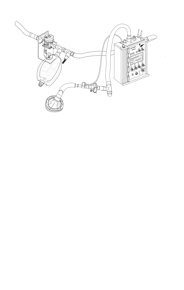
the draw-over system is shown in Figure 9-1.
Draw-over vaporizer.
ο Currently fielded model: Ohmeda Portable Anesthesia
Complete (PAC).
ο Demand type system (unlike the plenum systems in
hospital-based ORs).
♦ When the patient does not initiate a breath or the self-
inflating bag is not squeezed, there is no flow of gas.
No demand equals no flow.
ο Temperature-compensated flow-over in-line vaporizer.
9.9
Emergency War Surgery
150
Fig. 9-1. Draw-over apparatus in combination with the ventilator.
ο Optimal oxygen conservation requires a larger reservoir
(oxygen economizer tube [OET]) than is described in the
operator’s manual — a 3.5 ft OET optimizes FIO
2
ο May be used with spontaneous or controlled ventilations.
ο Bolted-on performance chart outlines dial positions for
some commonly used anesthetics (eg, halothane, isoflu-
rane, enflurane, and ether). Ether is highly flammable;
use extreme care.
Ohmeda UPAC Draw-Over Apparatus in Combination With
the Impact Uni-Vent Eagle 754 Portable Ventilator:
Currently, there is no mechanical ventilator specifically
designed for use with the UPAC draw-over apparatus, but
use with various portable ventilators has been studied in both
the draw-over and push-over configuration.
ο Adding the ventilator frees the anesthesia provider’s hands
while providing more uniform ventilation and more
consistent concentrations of the inhalational anesthetic
agent.
9.10
Anesthesia
ο The draw-over configuration places the ventilator distal
to the vaporizer, entraining ambient air and vapor across
the vaporizer in the same manner as the spontaneously
breathing patient. Do not attach a compressed source of
air to the Impact Uni-Vent Eagle 754 in this configuration
because the Uni-Vent Eagle 754 will preferentially deliver
the compressed gases and will not entrain air/anesthetic
gases from the UPAC draw-over.
ο The push-over configuration places the ventilator proximal
to the vaporizer, effectively pushing entrained ambient air
across the vaporizer and then to the patient.
The Impact Uni-Vent Eagle 754 portable ventilator (Figure 9-1)
is not part of the UPAC apparatus but is standard equipment
for the US military. It has been used in combination with the
Ohmeda UPAC Draw-Over Apparatus.
ο
The air-entrainment (side intake) port is used to create the
draw-over/ventilator combination.
♦ The side intake port of the ventilator contains a
nonreturn valve preventing back pressure on the
vaporizer which could result in erratic and inconsistent
anesthetic agent concentrations.
ο
The patient air-outlet port on the ventilator also contains
a nonreturn valve, preventing back flow into the ventilator
from the patient side.
ο
Scavenging of waste gases can be accomplished by
attaching corrugated anesthesia tubing to either the outlet
port of the Ambu-E valve (induction circuit) or the
exhalation port of the ventilator tubing (ventilator circuit)
venting to the outside atmosphere.
ο
The following items are added to the circuit to improve this
UPAC/Impact Uni-Vent Eagle 754 ventilator combination:
♦ Small and large circuit adapters to aid in attachment of
various pieces.
♦ PALL Heat and Moisture Exchange Filter to conserve
heat and limit patient contact with the circuit.
♦ Accordion circuit extender to move the weight of the
circuit away from the patient connection.
♦ O2 extension tubing to attach supplemental O2.
9.11
Emergency War Surgery
ο Two separate circuits should be constructed for use with
the UPACTM/Uni-Vent Eagle 754 combination: one for
induction and spontaneous ventilation and the second for
controlled ventilation using the portable ventilator.
♦ This process can be complicated because switching
circuit components requires several disconnections and
reconnections, creating the potential for error. (Practice.)
Conventional plenum anesthesia machine.
ο Currently fielded models: Drager Narkomed and Magellan
2000.
ο Compact version of standard OR machines, with com-
parable capabilities.
9.12
Chapter 10
Infections
Introduction
All wounds incurred on the battlefield are grossly
contaminated with bacteria. Most will become infected
unless appropriate treatment is initiated quickly.
The battlefield environment is conducive to wound infection
due to
Absence of “sterile” wounding agents on the battlefield. All
foreign bodies (wounding projectile fragments, clothing, dirt)
are contaminated with bacteria.
High-energy projectile wounding (devitalized tissue,
hematoma, tissue ischemia).
Delay in casualty evacuation.
Diagnosis of a Wound Infection
The four “-ors:” dolor, rubor, calor, and tumor—pain and
tenderness, redness, warmth, and swelling.
Drainage or discharge, ranging from frank pus to the foul
“dishwater” discharge of clostridial infection.
Crepitus, radiographic evidence of soft-tissue gas, epidermal
blistering, and/or epidermal necrosis are the hallmarks of
necrotizing soft tissue infection, such as clostridial gas
gangrene or necrotizing fasciitis.
Systemic effects such as fever, leukocytosis, unexplained
tachycardia, or hypotension.
Confirm diagnosis by Gram stain and culture, if available,
and/or tissue biopsy.
10.1
Emergency War Surgery
Common Microorganisms Causing Battlefield Infections
Gram-positive cocci: staphylococci, streptococci, and
enterococci.
Gram-negative rods: Escherichia Coli, Proteus, and Klebsiella.
ο Pseudomonas, Enterobacter, Acinetobacter, and Serratia are
common nosocomial pathogens usually expected among
casualties who have been hospitalized for an extended
period, not those fresh off the battlefield.
Salmonella, Shigella, and Vibrio should be suspected in cases
of bacterial dysentery.
Anaerobic Gram-positive and Gram-negative rods: Clostridia,
Bacteroides, and Prevotella species.
Fungal species: Candida species should be suspected in casualties
hospitalized for prolonged periods, those malnourished or
immunosuppressed, or those who have received broad
spectrum antibiotics, adrenocortical steroids, or parenteral
nutrition. Empiric therapy should be considered in appropriate
patients with presumptive evidence of fungal infection.
The greatest threat of infection to the wounded battlefield
casualty is the development of clostridial myonecrosis (gas
gangrene), commonly due to Clostridium perfringens.
Common Patterns of Infection
Skin, soft tissue, muscle, and bone: Primarily due to
staphylococcal, streptococcal, and clostridial species. These
infections include wound abscess, cellulitis, septic arthritis,
osteomyelitis, necrotizing fasciitis, and gas gangrene.
Clostridium tetani can enter through any wound—even
minor burns and corneal abrasions. Prophylaxis is required
to prevent tetanus toxemia.
Intracranial: Meningitis, encephalitis, and abscess,
commonly due to staphylococci and Gram-negative rods,
which are difficult to treat due to the impervious nature of
the meninges to common antibiotics.
10.2
Infections
Orofacial and neck: Gram-positive cocci and mouth
anaerobes, generally responsive to surgery and clindamycin.
Thoracic cavity: Empyema (usually staphylococcal) and
pneumonia (Staphylococcus, Streptococcus, Pseudomonas),
especially among those on prolonged mechanical ventilation
or those casualties prone to aspiration (polymicrobial).
Intraabdominal: Include posttraumatic or post-operative
abscess, and peritonitis due to Enterococcus, Gram-negative
rods, and anaerobic bacilli. Clostridium difficile is often
responsible for a potentially severe diarrheal colitis that
occurs following the administration of even one dose of
antibiotic.
Systemic sepsis: A syndrome caused by a bloodborne or
severe regional infection resulting in a global inflammatory
response (fever, leukocytosis, tachycardia, tachypnea, and
possibly hypotension).
ο A similar inflammatory response without infection can be
caused by a focus of retained necrotic tissue, or the mere
act of sustaining severe trauma.
ο Culprit microorganisms will not be recovered in all cases
of sepsis syndrome.
ο Although typically associated with Gram-negative
organisms, any bacterial or fungal agent can cause sepsis.
Prompt surgical debridement is the cornerstone of
prophylaxis/treatment of war wound infections.
Treatment
General Principles
Surgical and antibiotic treatment should begin early and be
repeated in the prophylaxis of war wound infection.
Optimally, surgical debridement should be achieved within
6 hours of injury.
Following initial exploration and debridement, the wound
should be sufficiently irrigated to ensure all dead material,
bacterial contamination, and foreign material has been
washed from the wound.
10.3
Emergency War Surgery
Excessive irrigation, especially under pressure, should be
avoided, because this can dilute the body’s natural immune
cellular defenses and contribute to bacteremia.
The skin is left open, and a lightly moistened sterile gauze
dressing is applied.
Antibiotics should be started ASAP after wounding, then
continued for 24 hours, depending on the size, extent of
destruction, and degree of contamination of the wound.
ο If time from wounding to initiation of antibiotics is > 6
hours, or time from wounding to surgery is > 12 hours,
give antibiotics using regimen for established infection.
The choice of empiric antibiotic is dependent on the part of
the body injured (Table 10-1).
Once a battlefield wound has become infected, treatment is
two-fold—surgical and medical.
ο Surgical strategy remains the same: Open the wound, remove
infected and necrotic tissue, and inspect for foreign material.
ο Drainage is generally employed in abscess cavities to
prevent premature closure and reformation.
ο Empiric broad-spectrum antibiotic therapy is initiated
against likely pathogens and continued for 7 to 10 days.
ο Ideally, obtain cultures and tailor therapy to cover the
actual pathogens recovered on Gram stain and culture.
Routine bacteriology is often not available in forward
medical facilities.
ο Because Bacteroides and Clostridia are difficult to culture,
tailor antibiotic therapy to cover these organisms.
ο If the debrided wound still has possibly ischemic tissue or
retained foreign material, the patient is returned to the OR
every 1 to 2 days for redebridement, until absolute
assurance of healthy, clean tissue is achieved.
Specific Infections
Tetanus.
ο Battlefield wounds are “tetanus-prone” due to high levels
of contamination with Clostridium tetani.
ο Bacteria grow anaerobically and release a CNS toxin that
results in muscle spasm, trismus, neck rigidity, and back
arching.
10.4
Table 10-1. Empiric Antibiotic Coverage for War Injuries.
Site of Injury
Empiric Antibiotic
Covered Organisms
Cranium/penetrating
Ancef/Vanc + Flagyl
Gram positives + anaerobes
injury
brain injury
Maxillofacial
Ancef + clindamycin
Gram positives + anaerobes
Neck
Ancef
Skin flora
Chest
Ancef
Skin flora
Abdomen
Liver
Fluoroquinolone/2nd generation
Gram negatives, gram positives,
cephalosporin
+ anaerobes
Gastrointestinal tract
Carbapenam/penicillin (Zosyn)
“
with gross contamination
Gastrointestinal tract
2nd generation cephalosporin
“
without gross contamination
Genitourinary
Aminoglycoside + 2nd generation
“
cephalosporin
Spleen
2nd generation cephalosporin +
“
fluoroquinolone + immunize splenectomy
patients later for encapsulated organisms
Pelvic
With gastrointestinal injury
Carbepenam or combo penicillin
Gut flora + anaerobes
No gastrointestinal injury
2nd generation cephalosporin
Skin organisms
Extremity
Soft tissue only
Ancef or 2nd generation cephalosporin
+ aminoglycoside
Bone/vascular involvement
2nd generation cephalosporin +
aminoglycoside and fluoroquinolone
Treat gross contamination of any wound with debris from uniforms and the environment with broad spectrum Gram-negative and
anaerobic coverage regardless of area of injury, eg, Ancef + penicillin + gentamicin; or Unasyn alone.
Emergency War Surgery
ο
In addition to surgical debridement of war wounds,
additional prophylactic measures for tetanus-prone
wounds include
♦ Administration of 0.5ml IM of tetanus toxoid if prior
tetanus immunization is uncertain, less than three doses,
or more than five years since last dose.
♦ Administration of 250-500 units IM of tetanus immune
globulin in a separate syringe and at a separate site from
the toxoid if prior tetanus immunization is uncertain
or less than three doses.
ο
Treatment for established tetanus includes
♦ IV antibiotics (penicillin G, 24 million U/d; or
doxycycline, 100 mg bid; or metronidazole, 500 mg q6h
for 7 days).
♦ Tetanus immune globulin.
♦ Wound debridement as needed.
♦ IV diazepam to ameliorate the muscle spasm.
♦ Place patient in a dark, quiet room free of extraneous
stimulation.
♦ May warrant endotracheal intubation, mechanical
ventilation, and neuromuscular blockade.
Soft-tissue infections.
ο Cellulitis is manifested by localized skin erythema, heat,
tenderness, and swelling or induration.
♦ Treatment: IV antibiotics against streptococcal and
staphylococcal species (IV nafcillin, cefazolin or, in the
penicillin-allergic patient, clindamycin or vancomycin).
ο Post-operative wound infections become evident by
wound pain, redness, swelling, warmth, and/or foul or
purulent discharge, with fever and/or leukocytosis.
♦ Treatment: Open the wound, drain the infected fluid,
and debride any necrotic tissue present.
♦ The wound is left open and allowed to close via
secondary intention.
ο Necrotizing soft tissue infections are the most dreaded
infections resulting from battlefield wounding. These
include clostridial myonecrosis (gas gangrene) and
polymicrobial infections caused by Streptococcus,
Staphylococcus, Enterococcus, Enterobacteriaceae, Bacteroides,
and Clostridia.
10.6
Infections
♦ The organisms create a rapidly advancing infection
within the subcutaneous tissues and/or muscle by
producing exotoxins that lead to bacteremia, toxemia,
and septic shock.
♦ All layers of soft tissue can be involved, including skin
(blistering and necrosis), subcutaneous tissue
(panniculitis), fascia (fasciitis), and muscle.
♦ Clinical manifestations begin locally with severe pain,
crepitus, and with clostridia, a thin, brown, foul-
smelling discharge.
♦ The skin may be tense and shiny, showing pallor or a
bronze color.
♦ Systemic signs include fever, leukocytosis, mental
obtundation, hemolytic anemia, and hypotension,
progressing rapidly to multiple organ failure and death
in untreated or under-treated cases.
♦ The diagnosis is made by history of severe unexpected
wound pain combined with palpable or radiographic
soft tissue gas (air in subcutaneous tissue and/or
muscle).
♦ Absence of soft-tissue gas does not exclude diagnosis
of necrotizing infection.
♦ Treatment is surgical, including early, comprehensive,
and repeated (every 24-48 hours) debridement of all
dead and infected tissue, combined with antibiotics.
♦ Excision of affected tissue must be as radical as
necessary (including amputation or disarticulation) to
remove all muscle that is discolored, noncontractile,
nonbleeding, or suspicious.
♦ Identification of causative organisms often problematic:
Treatment must be aimed at all possible organisms.
♦ IV antibiotic therapy.
♦ Clindamycin, 900 mg q8h; plus penicillin G, 4 million
U q4; plus gentamicin, 5-7 mg/kg qd.
◊ As a substitute for clindamycin: metronidazole, 500
mg q6h.
◊ As a substitute for penicillin: ceftriaxone, 2.0 g q12h,
or erythromycin 1.0 g q6h.
◊ As a substitute for gentamicin: ciprofloxacin, 400 mg
q12h.
10.7
Emergency War Surgery
♦ Alternative regimen: penicillin G, 4 million U q4h plus
imipenem, 500 mg q6h.
Intraabdominal infections.
♦ Prevention.
♦ Regimens (start ASAP, continue x 24 hours post-op).
◊ Single agent: cefotetan 1.0 g q12h, or ampicillin/
sulbactam, 3 g q6h, or cefoxitin, 1.0 g q8h.
◊ Triple agent: ampicillin 2 g q6h; plus anaerobic coverage
(metronidazole, 500 mg q6h; or clindamycin, 900 mg
q8h); plus gentamicin 5-7 mg/kg qd.
♦ Established intraabdominal infection (peritonitis or
abscess).
♦ Same regimen as above, except continue for 7 to 10 days.
♦ Drain all abscesses.
Pulmonary infections.
♦ Empyema (generally Streptococcal) following penetrating
thoracic trauma is typically due to contamination from
the projectile, chest tubes, or thoracotomy.
♦ Diagnosis: Loculations, air/fluid levels on radiograph,
pleural aspirate.
♦ Treatment.
◊ Chest tube initially, and thoracotomy if unsuccessful.
◊ Cefotaxime, or ceftriaxone, or cefoxitin, or imipenem.
♦ Pneumonia is most frequently due to aspiration (eg,
patients with head injury) and prolonged mechanical
ventilation.
♦ The diagnosis is made through radiograph finding of a
new pulmonary infiltrate that does not clear with chest
physiotherapy, combined with
◊ Fever or leukocytosis.
◊ Sputum analysis showing copious bacteria and
leukocytes.
♦ Empiric therapy is directed toward likely pathogens.
◊ Aspiration: Streptococcal pneumonia, coliforms, and
oral anaerobes are likely. IV antibiotics such as
ampicillin/sulbactam, clindamycin, or cefoxitin have
proven effective.
◊ Ventilator-associated pneumonia: staphylococcus,
Pseudomonas, and other nosocomial Enterobacteriaceae.
Broad coverage is best with such agents as imipenem,
10.8
Infections
ciprofloxacin, vancomycin, and/or ceftazidime, plus
an aminoglycoside.
Systemic Sepsis
Sepsis can be defined as infection combined with a prolonged
systemic inflammatory response that includes two or more of
the following conditions.
Tachycardia.
Fever or hypothermia.
Tachypnea or hyperventilation.
Leukocytosis or acute leukopenia.
Progression to septic shock is manifest by systemic
hypoperfusion: profound hypotension, mental obtundation, or
lactic acidosis. Treatment is a three-pronged approach:
Identify and eradicate the source.
Broad-spectrum intravenous antibiotics for the most likely
pathogens.
ICU support for failing organ systems, such as cardiovascular
collapse, acute renal failure, and respiratory failure.
It is often difficult to identify the source of sepsis, but it is the
most important factor in determining the outcome. Potential
sources of occult infection include
An undrained collection of pus such as a wound infection,
intraabdominal abscess, sinusitis, or perianal abscess.
Ventilator-associated pneumonia.
Urinary tract infection.
Disseminated fungal infection.
Central intravenous catheter infection.
Acalculous cholecystitis.
Intensive care support for sepsis involves vigorous resuscitation
to restore perfusion to prevent multiple organ dysfunction.
This requires optimization of hemodynamic parameters
(pulmonary artery occlusion pressure, cardiac output, and
oxygen delivery) to reverse anaerobic metabolism and lactic
acidosis. Endpoints of resuscitation, such as urine output,
base deficit, and blood lactate levels guide successful
treatment. Until the source for sepsis is identified and actual
pathogens isolated, empiric therapy with broad-spectrum
intravenous antibiotics is warranted. Suitable regimens might
include
10.9
Emergency War Surgery
Imipenem, 500 mg q6h.
Piperacillin and tazobactam (Zosyn), 3.375 g q6h; or
ceftazidime, 2.0 g q8h; or cefepime, 2.0 g q12h; PLUS
gentamicin, 5-7 mg/kg qd (based on once-daily dosing
strategy and no renal impairment); or ciprofloxacin, 400 mg
q12h.
Addition of vancomycin, 1.0 g q12h if methicillin-resistant
Staphylococcus aureus is a likely pathogen.
Addition of linezolid, 600 mg q12h if vancomycin-resistant
enterococcus (VRE) is a likely pathogen.
Conclusion
Battlefield casualties are at high risk for infection. In particular,
war wounds are predisposed to infection due to environmental
conditions on the battlefield, devitalized tissue, and foreign
bodies in the wound. The key to avoiding wound infection is
prompt and adequate wound exploration, removal of all foreign
material, and excision of all dead tissue. All battlefield wounds
and incisions should have the skin left open. Antibiotics play
an adjunctive role in the prophylaxis of wound and other
infections in the battlefield MTF. Knowledge of likely pathogens
for particular infections and sites, as well as optimal antibiotics
to eradicate those pathogens (Table 10-2), will aid the battlefield
clinician in averting and treating infections.
10.10

Table 10-2. Spectrum and Dosage of Selected Antibiotic Agents.
Agent
Antibacterial Spectrum
Dosage
Penicillin G
Streptococcus pyogenes, penicillin-sensitive Streptococcus pneumonia,
4 mil U IV q4h
clostridial sp
Ampicillin
Enterococcal sp, streptococcal sp, Proteus, some E coli, Klebsiella
2 g IV q6h
Ampicillin/
Enterococcal sp, streptococcal sp, Staphylococcus,* E coli,
3 g IV q6h
sulbactam
Proteus, Klebsiella, Clostridial sp, Bacteroides/Prevotella sp
Nafcillin
Staphylococcal sp,* streptococcal sp
1 g IV q4h
Piperacillin/
Enterococcal sp, streptococcal sp, Staphylococcus,* E coli, Pseudomonas
3.375 g IV q6h
tazobactam
and other enterobacteriaceae, clostridial sp, Bacteroides/Prevotella sp
Imipenem
Enterococcal sp, streptococcal sp, Staphylococcus,* E coli, Pseudomonas
500 mg IV q6h
and other enterobacteriaceae, clostridial sp, Bacteroides/Prevotella sp
Cefazolin
Staphylococcal sp,* streptococcal sp, E coli, Klebsiella, Proteus
1.0 g IV q8h
Cefoxitin
Staphylococcal sp,* streptococcal sp, E coli and similar enterobacteriaceae,
1.0 g IV q6h
clostridial sp, Bacteroides/Prevotella sp
Ceftazidime
Streptococcal sp, E coli, Pseudomonas and other enterobacteriaceae
2.0 g q8h
Ceftriaxone
Streptococcal sp, staphylococcal sp,* Neisseria sp, E coli and most
2.0 g q12h
enterobacteriaceae (NOT Pseudomonas), clostridial sp
Ciprofloxacin
E coli, Pseudomonas and other enterobacteriaceae
400 mg q12h
Gentamycin
E coli, Pseudomonas and other enterobacteriaceae
5-7 mg/kg qd (based on once-
daily dosing strategy and no
renal impairment)
Vancomycin
Streptococcal, enterococcal, and staphylococcal species (including MRSA;
1.0 g q12h
not VRE)
Erythromycin
Streptococcal sp, clostridial sp
0.5-1.0 g q6h
Clindamycin
Streptococcus sp, Staphylococcus sp,* clostridial sp, Bacteroides, and Prevotella sp
900 mg q8h
Metronidazole
Clostridial sp, Bacteroides and Prevotella sp
500 mg q6h
*Not methicillin resistant Staphylococcus aureaus (MRSA)
Dosage and dosage intervals are average recommendations. Individual dosing may vary.
Chapter 11
Critical Care
Introduction
Each battlefield ICU should have a dedicated intensive care
physician, due to the severity and lethality of blast and high-
velocity wounds, and the need for ongoing resuscitation of
casualties requiring damage control.
Damage control is the initial control of hemorrhage and
contamination followed by intraperitoneal packing and rapid
closure, then resuscitation to normal physiology in the
intensive care unit and subsequent definitive re-exploration.
This places large logistic requirements on the ICU. This may
include rewarming, large-volume resuscitation, blood
products, vasoactive drugs, and mechanical ventilation.
The ICU physician should observe the following guidelines:
Reexamine (possibly, retriage) the patient, using detailed
primary and secondary surveys, with attention to the “ABCs,”
potential life-threatening injuries, and other injuries missed
during the ER and OR phases of resuscitation (tertiary survey).
Trust no one’s examination before your own because the
patient’s condition may have changed, or prior examinations
may be inaccurate or incomplete.
Provide necessary available monitoring of physiology, with
periodic assessment of pain control, level of consciousness,
and intake and output.
Resuscitate from shock, using appropriate endpoints.
Provide organ-specific support, as is done for CNS injury,
pulmonary failure, cardiovascular collapse, and renal
dysfunction.
11.1
Emergency War Surgery
Ensure adequate pain control.
ο Use IV (not IM) narcotic agents in sufficient doses to
alleviate pain.
ο Patients on mechanical ventilation require both narcotics
(morphine, fentanyl) and sedatives (propofol, lorazepam,
midazolam).
Prepare the patient for transport out of theater.
Important caveats for the intensivist.
ο “Patients don’t often suddenly deteriorate; healthcare
providers suddenly notice!”
ο The organ system approach, in which each organ system
in turn is addressed in a mini-SOAP format, ensures that
each of the body’s physiologic systems is addressed in a
complete, comprehensive, and integral fashion.
ο The systemic inflammatory response (SIRS) is a common
metabolic sequela of severe injury, not always associated
with infection.
Fever or leukocytosis should prompt a thorough search for
infection. Antibiotic discipline must be enforced, saving these
medications for short-course prophylaxis, documented
infection, or empiric treatment of rapid deterioration due to
sepsis.
Resuscitation From Shock
Shock can be defined as an acute state of cardiovascular
insufficiency resulting in life-threatening global
hypoperfusion. Hemorrhagic shock is the most common form
of shock following major trauma. Therefore, initial efforts
should be directed toward correction of hypovolemia.
Hypoperfusion implies inadequate delivery of oxygen to the
body’s cells. Oxygen delivery is a function of cardiac
performance, arterial hemoglobin content, and arterial oxygen
saturation. All attempts to correct shock involve optimizing
these three variables.
11.2
Critical Care
Shock resuscitation is approached in two phases, based on
endpoints of resuscitation:
ο In the first phase, resuscitate to a mean arterial pressure
of > 60 mm Hg, a urine output of 0.5 cc/kg/h (at least 30
ccs/h), and arterial oxygen saturation of > 92%.
ο Pursue endpoints aggressively to eliminate hypoperfusion,
ideally within 1 hour (see Chapter 7, Shock and
Resuscitation).
ο In the second phase, resuscitation is continued primarily
with fluid, to eliminate metabolic acidosis (restore lactate
to normal) within 24 hours.
ο The resuscitative fluid of choice is a warmed, balanced
crystalloid solution (normal saline or lactated Ringer’s)
and is preferable to colloid.
ο Rate of infusion for resuscitation should be 500 mL to 1,000
mL bolus over 15-20 minutes and repeated as necessary.
ο After 3 L of crystalloid, blood products should generally
follow at similar rates.
Vasopressor agents should only be considered for achieving
minimal acceptable blood pressure after fluid boluses and
confirmation of adequate intravascular volume.
ο Dopamine, norepinephrine, and phenylephrine are the
preferred vasoactive agents, starting in the lower dose range.
ο Dobutamine should only be considered for demonstrated
cardiac dysfunction, which may be seen in sepsis, the
elderly, or myocardial infarction (MI).
Specific Organ Systems
Traumatic Brain Injury/CNS
Transient hypoxemia or hypotension in the patient with
significant traumatic brain injury doubles the probability of
death or poor neurologic outcome. The goal of treatment is to
maintain cerebral perfusion pressure (CPP) and oxygenation.
11.3
Emergency War Surgery
Identify potential intracranial surgical lesions for possible
emergent craniotomy.
Prevent hypoxemia: Maintain O2 sat > 92%, PaO2 > 100, and
intubate for GCS < 8.
Prevent hypotension.
ο Maintain SBP > 100 mm Hg, MAP > 80.
♦ MAP = DBP + 1⁄3 (SBP - DBP).
Prevent, monitor, and treat intracranial hypertension.
ο Maintain intracranial pressure (ICP) = 5-15 mm Hg.
ο Maintain CPP = 70-90 mm Hg.
CPP = MAP - ICP
Measures to treat intracranial hypertension include:
ο Elevation of head of bed 30° may be helpful.
ο Recognize that high levels of PEEP may raise ICP.
ο Control serum osmolarity.
♦ Normal saline is the preferred IV solution.
♦ Check serum sodium twice daily, and keep in the range
of 145-150 mEq/dL.
♦ IV mannitol (not in anuric patients), 0.25-1.0 g/kg,
every 6-8 hours to keep serum osmolarity optimal.
ο Control PaCO2.
Hypercarbia should always be prevented. Modest
therapeutic hyperventilation may be used (PaCO2 30-35 mm
Hg) for brief periods in the deteriorating patient.
♦ Beneficial effects of hyperventilation/hypocarbia must
be balanced: it reduces ICP through vasoconstriction,
but also reduces cerebral blood flow.
♦ Prophylactic hyperventilation should not be used.
ο Removal of cerebrospinal fluid by placement of an
intraventricular catheter.
ο Barbiturates have unproven benefit but may be considered
in extreme cases.
ο Craniotomy with bone and brain removal is a drastic,
lifesaving procedure of last resort in the moribund patient.
Steroids have no role in traumatic brain injury treatment.
11.4
Critical Care
ο Avoid hyperthermia, because this raises ICP.
General Considerations.
ο Appropriate precautions should be taken (H2 blocker,
heparin, and oral care) to prevent development of stress
gastritis, deep venous thrombosis, and aspiration
pneumonitis.
ο If coagulopathy develops, use blood products as necessary
to correct an elevated prothrombin time.
ο Prevent and aggressively treat pain, agitation, shivering,
and fever to avoid increased cerebral metabolism and
oxygen consumption.
ο Hyperglycemia has an adverse effect on outcome and
should be monitored and treated aggressively to keep
glucose levels between 100-150 mg/dL.
ο Seizure prophylaxis. Phenytoin/phosphenytoin should be
administered to therapeutic levels in the penetrating head-
injured patient, and to blunt head-injury patients with
seizure.
Pulmonary System and Ventilators
General Considerations
Supplemental oxygen in the early phase of resuscitation is
imperative. The maximum fraction of inspired oxygen (FIO2)
delivered by:
ο Nasal cannula approaches 0.35.
ο Venturi mask is 0.50.
ο Non-rebreathing reservoir mask approaches 0.90.
Monitoring: May include portable chest radiographs,
periodic ABGs, regular assessments of level of sedation,
airway pressures, and functioning of ventilator alarms.
Airway Considerations
Indications for endotracheal intubation and mechanical
ventilation include:
ο Airway obstruction due to trauma, edema, excess
secretions.
ο Apnea.
11.5
Emergency War Surgery
ο Excessive work of breathing (eg, flail chest), as indicated
by accessory muscle use, fatigue, diaphoresis, or tachypnea
when respiratory failure is imminent.
ο Decreased level of consciousness: GSC < 8.
ο Hypoxia: SaO2 < 90%, PaO2 < 60 mmHg on FIO2 > 50%.
ο Hypercarbia: PaCO2 > 60 mm Hg acutely (lower threshold
with tachypnea).
ο Shock.
ο Caution: Patients not meeting the above criteria may still
require airway protection and mechanical ventilation
preceding prolonged transport.
Field Ventilator.
ο
Impact Uni-Vent Eagle 754.
♦ Basic settings.
◊
Turn the ventilator on and set the mode using the
Mode Selector Switch (lower right). Most patients
can be well ventilated using SIMV (synchronized
intermittent mandatory ventilation).
◊
Set FIO2 using the Air/Oxygen Mixer Control, above
the Mode Selector Switch. Generally, ICU patients
should be started at an FIO2 of 1.0, and weaned as
appropriate to a level of 0.40.
◊
Set minute ventilation using Tidal Volume and
Ventilation Rate controls. The tidal volume is set at
6-10 mL/kg. Initial rate is set at 10-14 breaths/
minute and titrated to normalize PaCO2.
◊
Set positive end-expiratory pressure (PEEP) using the
PEEP Control located on the upper left of the control
panel. Initial PEEP is usually set at 5 cm H2O. Higher
values can be set in severe respiratory failure such
as adult respiratory distress syndrome (ARDS),
although generally not higher than 15 cm H2O.
Summary of typical initial settings for mechanical
ventilator: FIO2 1.0, SIMV mode, rate 12, tidal volume 800
mL, PEEP 5 cm H2O.
11.6
Critical Care
ARDS
ARDS can start within days of injury, and should be suspected
in any casualty with:
ο Acute hypoxemia (PaO2/FIO2 ratio < 200).
ο Progressive fall in pulmonary compliance (stiff lungs,
increasing airway pressures).
ο Bilateral alveolar infiltrates on chest radiograph, with no
clinical evidence of volume overload.
Mechanical Ventilation Priorities in ARDS
Maintain patient analgesia and sedation to prevent agitation
and ventilator/patient asynchrony.
Keep SaO2 > 90% by increasing FIO2 and/or PEEP (maximum
15-18 cm H2O).
Avoid prolonged FIO2 > 0.60 due to O2 toxicity.
Avoid respiratory acidosis. Keep PaCO2 35-45 mm Hg, and
arterial blood pH > 7.25.
Keep peak inspiratory pressure (PIP) < 40 cm H2O to prevent
iatrogenic pneumothorax and destruction of normal lung tissue.
ο Decrease tidal volume to 5-7 mL/kg.
ο Increase ventilator rate.
ο If other measures are unsuccessful, allow permissive
hypercapnia by accepting a respiratory acidosis (PaCO2
55-70 mm Hg). Use bicarb to maintain pH > 7.2.
Respiratory acidosis is less dangerous than ventilator-induced
lung injury caused by high PIP and high tidal volumes.
Cardiovascular System
The patient who exhibits cardiovascular deterioration after
a period of apparent stability should be evaluated to rule out
the following:
ο Hypoxia or loss of airway.
ο Tension pneumothorax.
ο Recurrent bleeding from sites of injury or surgery.
ο Cardiac tamponade or direct myocardial injury.
11.7
Emergency War Surgery
ο Tachyarrhythmia.
ο Fluid loss due to “third-spacing,” burns, fever, diarrhea,
or vomiting.
ο Undiagnosed injury: intestinal injury, pancreatitis, or
infection.
ο Vasodilatation due to spinal shock, epidural anesthesia/
analgesia, and sepsis.
ο Side effect from medication.
ο GI bleeding.
ο Pulmonary embolus.
ο Abdominal compartment syndrome.
ο Excessive airway pressures from mechanical ventilation
can directly decrease cardiac ventricular function and
decrease venous preload.
Management.
ο
Support cardiovascular system by monitoring end-organ
perfusion (urine output, capillary refill) and using four
parameters of hemodynamic performance:
♦ Preload (Best index: pulmonary capillary wedge
pressure - PCWP).
♦ Afterload (systemic vascular resistance [SVR] = [MAP
- CVP]/ CO • 80).
♦ Heart rate.
♦ Cardiac contractility (best index: stroke volume; SV =
CO/h).
♦ “Make do” with the best information available—use
CVP when PA catheter unavailable.
♦ For hypovolemia and cardiovascular instability due to
sepsis:
◊ Assure adequate preload by volume repletion before
adjusting other variables (eg, adding inotropes for
low cardiac output).
◊ In other states of cardiovascular instability, the
variable manipulated is the one indicative of the
major problem.
Sinus tachycardia may be a sign of an underlying problem
(eg, hypoxia, hypovolemia, infection, or pain). Seek and
treat the primary problem, not the tachycardia.
11.8
Critical Care
Myocardial ischemia/infarction (MI) is an uncommon
battlefield problem.
ο Suspicion is aroused when the patient exhibits angina-like
chest pain or unexplained cardiac instability (arrhythmias
or hypotension).
ο The diagnosis of an acute myocardial event is made by
the presence of ST segment elevation or depression on 12-
leak ECG and/or an abnormal elevation of serum markers
of myocardial injury (myoglobin [MB] fraction of creatine
phosphokinase, Troponin I).
Emergency treatment for MI.
ο Supplemental O2.
ο Morphine for pain, rest.
ο Aspirin 325 mg tablet chewed and swallowed (then one
tablet PO qd).
ο NTG SL (0.4 mg tablet every 5 minutes until pain relieved,
maximum 3 doses) or IV infusion depending on severity
of condition.
ο Beta-blocker such as metoprolol (5-15 mg IV slowly q6h
or 50-100 mg PO q12h) or atenolol (50-100 mg PO) on
diagnosis and daily.
ο As resources and patient condition permit for MI with
diagnostic ECG: Optimal therapy would also include,
within 6 hours of symptoms, IV heparin and a thrombolytic
such as tissue plasminogen activator.
Renal System and Electrolytes
Monitor urine output, blood urea nitrogen (BUN), serum
creatinine, and serum electrolytes.
Acute renal failure (ARF) is manifested by oliguria (< 0.5 cc/
kg/h) and a rise in BUN and creatinine. The most frequent
causes for ARF are:
ο Hypovolemia.
ο Acute tubular necrosis (ATN) due to:
♦ Hypovolemia.
♦ Sepsis, IV contrast agents, aminoglycoside antibiotics,
or NSAIDs.
ο Crush, massive, soft-tissue injury or compartment syndrome,
with resultant rhabdomyolysis and myoglobinuria.
11.9
Emergency War Surgery
♦ In ARF due to rhabdomyolysis, consider administering
large volumes of IV fluid (300-800 mL/h), combined
with 50 mEq NaHCO3/L, to alkalinize the urine with
the goal of achieving a urine output of 2.0 cc/kg/h.
♦ Bilateral renal or ureteral trauma.
Two hours of oliguria (< 20cc/h) in an ICU patient (almost
always due to inadequate resuscitation) warrants aggressive,
immediate action.
Algorithm for hemodynamically stable ICU patient with
profound oliguria or anuria:
ο
Irrigate or replace Foley catheter to ensure function.
ο
After ensuring no signs of intravascular volume overload
(diffuse pulmonary crackles, S3 heart sound), administer
bolus of 1-2 L IV saline over 30 minutes.
ο
Review medication list and medical history to elicit potential
factors causing ARF; stop any agents that could contribute.
ο
Send any urine to lab with serum sample to calculate
fractional excretion of sodium (FENA) = (UNA • PCR)/(PNA
• UCR); FENA < 1.0 indicates prerenal cause (eg,
hypovolemia); FENA > 2.0 points to renal insult (ATN;
myoglobinuria) or postrenal cause (obstruction).
ο
Consider sonogram of kidneys to rule out bilateral renal
obstruction.
ο
Consider pulmonary artery catheter to optimize preload
(PCWP).
ο
Once PCWP > 16-18 mm Hg, and urine output minimal/
nonexistent, administer furosemide in escalating doses IV
bolus: 40, 80, 160, 240 mg max ( > 100mg = ototoxic).
Combine last dose with single dose 1.0 g chlorothiazide
IV, or administer 10 mg metolazone PO given 30 minutes
before last dose. Also consider furosemide drip or
metolazone drip.
If ineffective or if other complications of ARF occur, arrange
for dialysis as a temporizing renal support until spontaneous
renal recovery occurs. This means the physician must
optimize the casualty for transport out of theater, with special
attention to volume status, potassium, and acid base status.
11.10
Critical Care
ο Indications for dialysis in the casualty with ARF:
♦ Anuria beyond 8-12 hours.
♦ Hypervolemia.
♦ Hyperkalemia.
♦ Acidosis.
♦ Complications of uremia: mental status changes,
pericardial rub.
♦ Toxic levels of drugs/medications (eg, digoxin).
Hyperkalemia.
ο Verify again hyperkalemia (serum K > 6 mEq/L) and
serum pH.
ο Give IV calcium chloride, 10 mL of 10% solution over 5
minutes.
ο Give IV NaHCO3, 50 mEq over 5 minutes.
ο Give IV Dextrose (50g D50) 50 grams + 10 units regular
insulin IV over 10 minutes.
ο Recheck K+.
ο Give beta-agonist albuterol 10-20 mg over 15 minutes by
inhalation.
ο Consider enteral K-binding with enema of sodium
polystyrene sulfonate, 25-50 g, in sorbitol.
Hypokalemia: Treatment: 10-20 mEq KCL IV/hour in
monitored setting; difficult to treat hypokalemia unless
concomitant hypomagnesemia is first corrected.
Hypernatremia: Usually indicative of free water deficit. Water
deficit (L) = 0.6 • weight (kg) • [(measured serum Na)/
(normal serum Na of 140) - 1]. Half of this deficit should be
replaced over first 12-24 hours, and the remainder over the
next 1-2 days.
Hyponatremia: Indicative of excess of free water or
vasopressin (SIADH). Levels of serum sodium < 125 mEq/L
are associated with mental status changes or seizures.
Treatment should involve free water restriction or use of IV
normal saline, with goal of correction of sodium level no more
than 15 mEq/L over 24 hours, to prevent complication of
central pontine myelinolysis.
Hypophosphatemia: Phosphate is important as an energy
source, and should be repleted to level of 2.5 mg/dL with IV
KPO4 or NaPO4, 30 mMol over 1 hour.
11.11
Emergency War Surgery
Hyperphosphatemia (usually associated with ARF):
Phosphate levels over 6.0 mg/dL should be treated by enteral
binding agents, such as calcium acetate or sucralfate.
Hypomagnesemia: Administer 2 g magnesium sulfate IV in
solution over 60 minutes to goal of serum level of 2.0 mEq/dL.
Metabolic acidosis: Primarily lactic (most commonly due
to hypovolemia) and ketoacidosis. Neither should be treated
with sodium bicarbonate (it is contraindicated in lactic
acidosis). Sodium bicarbonate has very limited role in ICU
disorders: hyperkalemia, alkalinization of urine in
myoglobinuria, bicarbonate-responsive renal tubular acidosis
(RTA), and for massive gastrointestinal losses of bicarbonate
(profound diarrhea, enterocutaneous fistula).
Metabolic alkalosis: NG suction of stomach acid causes a
hypochloremic alkalosis, responsive to replacement of NG
losses with crystalloid. Excessive loop diuretic use can also
cause metabolic (contraction) alkalosis. If further diuresis is
needed, use a carbonic anhydrase inhibitor (acetazolamide
250 mg IV every 6 h) for 1-2 days.
Hematologic System
Most common coagulation disorder: dilutional coagulopathy.
ο Others include heparin-induced thrombocytopenia,
disseminated intravascular coagulation, coagulopathy due
to hypothermia or diffuse hepatic damage, and
thrombocytopenia.
ο Most require replacement transfusion of appropriate blood
products.
To prevent trauma-related deep venous thrombosis (DVT)
and pulmonary embolism, prophylactic measures
(subcutaneous heparin or sequential compression devices)
are required.
Gastrointestinal System and Nutrition
Prolonged shock can lead to GI dysfunction.
ο Stress gastritis: Increased risk of severe head injuries or
burns, mechanical ventilation, systemic anticoagulation
therapy, or sepsis. Prevention: sucralfate, histamine-2
receptor antagonist (eg, ranitidine) or a proton pump
inhibitor (eg, omeprazole).
11.12
Critical Care
ο Acalculous cholecystitis: Suspect with right upper quadrant
abdominal pain, abnormalities in liver function tests, or
fever/leukocytosis of unclear cause. Ultrasound shows
gallbladder inflammation with wall thickening or
pericholecystic fluid. Treatment: broad-spectrum antibiotics
and ultrasound-guided percutaneous drainage or operation.
ο Hepatic failure portends a dire prognosis. Initial signs
include hyperbilirubinemia, elevation of the prothrombin
time, hypoalbuminemia, profound hypoglycemia,
obtundation. Massive amounts of fresh frozen plasma are
required to prevent exsanguination from coagulopathy.
Nutrition can prove problematic in the battlefield ICU patient.
ο
Systemic inflammation induced by severe injury often
results in catabolism and protein wasting, making early
nutritional support imperative.
ο
Nutrition should commence within 24-48 hours of injury.
ο
Enteral feedings are superior to parenteral nutrition (TPN),
offering a lower infection rate and shorter ICU stays.
ο
The following goals serve to guide nutritional
management:
♦ Caloric requirement: 25-30 kcal/kg/d.
♦ Protein requirement: 1.0-1.5 g/kg/d.
♦ 30%-40% of total caloric intake per day should be as fat.
ο
Nutrition should include a balanced electrolyte solution
containing supplemental potassium, calcium, magnesium,
phosphate, multivitamins and trace elements (zinc, copper,
manganese, and chromium).
ο
The two most common problems associated with enteral
nutrition are diarrhea and aspiration.
♦ Aspiration can be associated with severe pneumonitis,
but can be prevented by:
◊ Keeping the head of the bed up.
◊ Feeding into the jejunum or duodenum rather than
the stomach.
◊ Checking gastric residuals every 4 hours (feedings
should be stopped if residual greater than 200 mL).
♦ Diarrhea can be alleviated by:
◊ Decreasing the osmolarity of the enteral solution.
◊ Adding fiber.
◊ Agents such as loperamide in small doses.
11.13
Emergency War Surgery
Immune System and Infections
Differential diagnosis of ICU infections.
ο Pneumonia (nosocomial or aspiration).
ο Central venous catheter infection - if considered, remove
catheter.
ο UTI.
ο Wound or soft-tissue infection.
ο Intra-abdominal abscess
(especially following
laparotomy).
ο Systemic fungal infection.
ο Sinusitis.
ο Acalculous cholecystitis.
ο Pancreatitis.
Prophylactic antibiotics.
ο A short course of prophylactic antibiotics (24-48 h) is
warranted after penetrating injury on the battlefield.
ο After this, antibiotics should be withheld unless a
documented infection is confirmed, or a severe deterioration
in clinical status suggestive of sepsis is encountered.
ο Sepsis warrants a short course of broad spectrum IV
antibiotics, but they must be stopped in 72 hours if no
microbiologic pathogens are confirmed by culture.
ο Fever and leukocytosis, by themselves, are not sufficient
justification for antibiotics.
Endocrine System
Hyperglycemia.
ο Control, to prevent ketoacidosis, hyperosmolar coma, and
intravascular volume loss due to osmotic diuresis.
ο The two most common causes are uncontrolled or
unrecognized infections and the use of TPN.
ο The best technique for control of hyperglycemia is a constant
IV infusion of insulin, usually 1.0-10 units per hour.
♦ Due to frequent problems with patient perfusion,
subcutaneous injections are less reliable in the ICU patient.
♦ Patients with profound hyperglycemia (serum glucose >
800 mg/dL) and volume depletion, due to osmotic
diuresis, should receive fluid resuscitation with crystalloid
11.14
Critical Care
before receiving insulin, to prevent further shifts in
intravascular volume as the glucose shifts intracellularly.
♦ Limit correction rate to 100 mg/dL per hour (700 mg/dL
takes 7 h to correct) and assess for resultant hypokalemia.
Corticosteroids are rarely indicated after major trauma.
ο There is no proven benefit to steroid treatment for closed
head injury or sepsis.
ο Steroids are indicated for proven adrenocortical deficiency
(a rare occurrence among battlefield casualties) and spinal
cord injury with neurologic deficit.
Musculoskeletal System
Monitor for the development of compartment syndrome,
vascular ischemia, and rhabdomyolysis.
Distal extremities should be assessed regularly for
neurovascular status: presence of pulses, sensation, motor
function, warmth, and skin color.
Preparation for Evacuation
Optimally, the combat casualty will be medically stabilized
before transport out of theater.
ο Native or mechanical airway is maintained.
ο Sufficient blood pressure, to allow organ perfusion, that
has been stable for at least 8 hours.
ο Both primary and secondary phases of shock resuscitation
have been completed.
ο All sources of bleeding have been identified and controlled.
ο Life-saving or definitive surgery not required for the next
24 hours.
Transfer out of a battlefield ICU requires a USAF Critical Care
Air Transport Team (CCATT) with physician-to-physician
and nurse-to-nurse communication to summarize condition
of the patient, operations performed, treatment being given,
and support required during flight (in particular, need for
oxygen, mechanical ventilation, suction, blood products, and
monitoring).
Copies of medical records, radiographs, 3 days of IV fluid,
and all medications should accompany the patient.
11.15
Chapter 12
Damage Control Surgery
Introduction
The traditional approach to combat injury care is surgical
exploration with definitive repair of all injuries. This approach
is successful when there are a limited number of injuries.
Prolonged operative times and persistent bleeding lead to the
lethal triad of coagulopathy, acidosis, and hypothermia,
resulting in a mortality of 90%.
Damage control is defined as the rapid initial control of
hemorrhage and contamination, temporary closure,
resuscitation to normal physiology in the ICU, and
subsequent re-exploration and definitive repair. This
approach reduces mortality to 50% in some civilian
settings.
What might increase the life and limb salvage rate in troops
in the field setting is the application of the damage control
concepts described above in patients with favorable
physiology.
Tactical Abbreviated Surgical Control (TASC).
ο Damage control techniques in a tactical environment.
ο Abbreviated, focused operative interventions for peripheral
vascular injuries, extensive bone and soft tissue injuries,
and thoracoabdominal penetrations in patients expected
to survive, instead of definitive surgery for every casualty.
ο This may conserve precious resources, such as time,
operating table space, and blood.
This TASC philosophy relies on further definitive surgical
care at the next echelon of care.
12.1
Emergency War Surgery
Damage control techniques should be considered in all multi-
system casualties at the onset of surgical therapy. When initially
rejected, reconsideration should occur when unexpected
findings are discovered or natural breaks in the surgical therapy
occur, following an initial decision to perform a definitive repair.
The goal of damage control is to restore normal physiology
rather than normal anatomy. It is used for the multiple injured
casualty with combinations of abdominal, vascular, genitou-
rinary, neurologic, orthopedic, and/or thoracic injury in three
separate and distinct phases:
1. Primary Operation and Hemorrhage Control - surgical
control of hemorrhage and removal of contamination;
laparotomy terminated, abdomen packed and temporary
closure; definitive repair is deferred.
2. Critical Care Considerations - normal physiology restored
in ICU by core rewarming, correction of coagulopathy, and
hemodynamic normalization.
3. Planned Reoperation - re-exploration to complete the
definitive surgical management or evacuation.
General Considerations
Philosophy of damage control is “a live patient above all else.”
ο Avoid hypothermia.
ο Rapidly achieve hemostasis.
ο Perform only essential bowel resections.
ο Close or divert all hollow viscus injuries, only performing
reconstruction at the second operation after the patient has
stabilized and can tolerate a prolonged operation.
When to employ damage control.
ο Use damage control in patients who are present with or at
risk for developing:
♦ Multiple life-threatening injuries.
♦ Acidosis (pH < 7.2).
♦ Hypothermia (temp < 34°C).
♦ Hypotension and shock on presentation.
♦ Combined hollow viscus and vascular or vascularized
organ injury.
♦ Coagulopathy (PT > 19 sec and/or PTT > 60 sec).
12.2
Damage Control Surgery
♦ Mass casualty situation.
ο Take into account ability to control hemorrhage, severity
of liver injury, and associated injuries.
ο Pack before massive blood loss (10-15 units of pRBCs) has
occurred.
ο Injuries that typically require damage control techniques.
♦ Upper abdominal injuries that are not isolated spleen
injuries (duodenal, large liver injuries, pancreas, and
so forth).
♦ Major penetrating pelvic trauma of more than one
system.
♦ Any retroperitoneal vascular injury.
To reiterate, damage control is practiced in three phases:
1. Primary operation and hemorrhage control.
2. Critical care resuscitation.
3. Planned reoperation.
Phase 1: Primary Operation and Hemorrhage Control
Phase 1 of damage control includes 5 distinct steps:
1. Control of hemorrhage.
2. Exploration to determine extent of injury.
3. Control of contamination.
4. Therapeutic packing.
5. Abdominal closure.
Control of hemorrhage/Vascular injury repair.
ο Control of hemorrhage is best done with ligation, shunting,
or repair of injured vessels as they are encountered.
ο The primary goal is hemorrhage control, not maintenance
of blood flow.
ο For the patient in extremis, clamping or shunting of major
vessels is recommended over repair.
♦ THINK: ligate/shunt ⇒ fasciotomy.
ο Additional methods of hemorrhage control include balloon
catheter tamponade of vascular or solid viscus injuries.
Exploration to determine extent of injury.
ο Damage control laparotomy.
♦ Rapidly achieve hemostasis.
12.3
Emergency War Surgery
♦ Perform only essential resections or pack solid organs
to diminish blood loss.
♦ Close or divert all hollow viscus injuries.
♦ Rapidly terminate the procedure to correct hypo-
volemia, hypothermia, and acidosis to prevent
coagulopathy.
♦ Perform definitive reconstruction only after the patient
has stabilized and can tolerate a prolonged operation.
Control of Contamination.
ο
Contamination control also proceeds as injuries are
encountered, utilizing clamps, primary repair or resection
without reanastomosis.
ο
With multiple enterotomies, if the area of injury represents
less than 50% of the length of the small bowel, a single
resection can be undertaken.
ο
At this stage of the operation, the surgeon must decide
whether or not to proceed with definitive repair of the
identified and controlled injuries. Careful communication
with the anesthesiologist is critical to this decision.
♦ If aggressive resuscitation has been successful in
maintaining normal temperature, coagulation, and acid
base status, then definitive repair may proceed.
♦ If any of these interrelated factors are abnormal, the
procedure should be terminated (contamination
controlled without reanastomosis) and the patient taken
to the ICU for further resuscitation.
♦ The presence and status of extra-abdominal injuries
needs to be taken into consideration when deciding how
much physiologic reserve the patient has left.
Therapeutic Packing.
ο Resuscitative vs Therapeutic Packing.
♦ Resuscitative packing is manual compression of the
bleeding site as an initial measure in controlling or
minimizing blood loss.
♦ Therapeutic packing provides long-term tamponade of
liver, pelvic, and retroperitoneal bleeding.
ο Do not use the “pack and peek” technique wherein the
liver is packed and the patient resuscitated; the packs are
removed to identify the source of bleeding, but rebleeding
12.4