Emergency War Surgery (2004) - page 10
Chapter 28
Burn Injuries
Introduction
Burns constitute between 5% and 20% of combat casualties
during conventional warfare, and are particularly common
during war at sea and combat involving armored fighting
vehicles. Even relatively small burns can be incapacitating, and
can strain the logistical and manpower resources of military
medical units. Optimal treatment currently results in salvage of
approximately 50% of young adults whose burns involve 80%
of the total body surface area (TBSA) or greater. Thus, in a battlefield
triage scenario, expectant care should be considered for patients
with burns that exceed 80%. Care can be delayed for those patients
with burns of 20% or less who are otherwise stable.
Point-of-Injury Care
The following are key steps in the first aid of burn patients:
Stop the burning process. Extinguish and remove burning
clothing, and remove the patient from a burning vehicle or
building. In an electrical injury, remove the patient from the
power source, while avoiding rescuer injury. Wash chemical
agents from the skin surface with copious water lavage.
Ensure airway patency, control hemorrhage, and splint
fractures.
Remove all constricting articles, such as rings, bracelets,
wristwatches, belts, and boots. However, do not undress the
patient unless the injury has been caused by a chemical agent,
in which case remove all contaminated clothing.
Cover the patient with a clean sheet and a blanket, if
appropriate, to maintain body temperature and to prevent
gross contamination during transport to a treatment facility;
special burn dressings are not required. Hypothermia is a
complication of large surface area burns.
Establish intravenous access through unburned skin if
possible, and through burned skin if necessary. Intraosseous
access is also acceptable.
28.1
Emergency War Surgery
Begin resuscitation with lactated Ringer’s solution (LR) or
similar solution, and continue during evacuation.
Dress white phosphorus-injured patients with saline-soaked
dressings to prevent reignition of the phosphorus by contact
with the air.
Primary Survey
Do not be distracted by the burn! The priorities of
management for burn casualties are the same as those
for other injured patients, with the addition of burn
pathophysiology.
The primary survey includes airway management (with
cervical spine control, if appropriate given the mechanism of
injury), diagnosis and management of any breathing
condition, rapid circulatory assessment, and hemorrhage
control. In the burn patient, special attention to exposure,
removal of clothing that continues to burn the victim, and
prevention of hypothermia are important.
Airway.
Inhalation injury may be manifested by stridor,
hoarseness, cough, carbonaceous sputum, dyspnea, and
so forth. It may cause airway obstruction at any time
during the first 2 days postburn.
Patients who may have sustained inhalation injury
should be closely observed in an intensive care unit, and
may be monitored without intubation if minimally
symptomatic.
Prior to transport, prophylactically intubate patients
having/or who have symptomatic inhalation injury.
Tubes, such as the orotracheal tube, should be
definitively secured with cloth ties (eg, umbilical tape).
Avoid adhesive tape.
ο Cervical spine injury is uncommon in burn patients, except
in those injured in explosions, high-speed vehicular accidents,
and falls, or by contact with high-voltage electricity.
28.2
Burn Injuries
ο Burns are a “distracting injury,” pain secondary to burns,
and the treatment of pain with narcotics, may make the
clinical diagnosis of spinal injury difficult.
Breathing.
ο Inhalation injury is more common in patients with
extensive cutaneous burns, a history of injury in a closed
space (eg, building or vehicle), facial burns, and at the
extremes of age.
ο Patients with major burns and/or inhalation injury require
supplemental oxygen, pulse oximetry, chest radiograph
and arterial blood gas measurement.
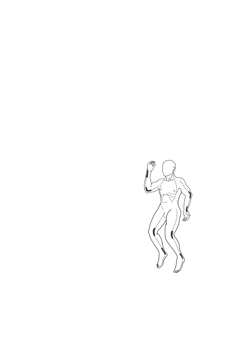
ο Circumferential burns of the chest may prevent effective
chest motion. If this occurs, perform immediate thoracic
escharotomy as a life-saving procedure to permit
adequate chest excursion (see Fig. 28-1).
ο Definitive diagnosis of lower airway injury requires
fiberoptic bronchoscopy.
Fig. 28-1.The dashed lines indicate the
preferred sites for escharotomy incisions.
The bold lines in the figure indicate the
importance of extending the incision over
involved major joints. Incisions are made
through the burned skin into the
underlying subcutaneous fat using a
scalpel or electrocautery. For a thoracic
escharotomy, begin incision in the
midclavicular lines. Continue the incision
along the anterior axillary lines down to
the level of the costal margin. Extend the
incision across the epigastrium as needed.
For an extremity escharotomy, make the
incision through the eschar along the mid-
medial or mid-lateral joint line.
ο Carbon monoxide (CO) poisoning causes cardiac and neurologic
symptoms. Patients with CO poisoning require 100% oxygen
for at least 3 hours or until symptoms resolve.
Circulation.
o Secure all cannulae (peripheral and central) with suture,
because tape will not adhere well.
28.3
Emergency War Surgery
ο Cuff blood pressure (BP) measurements may be inaccurate
in patients with burned or edematous extremities. Arterial
BP is preferred.
Estimation of Fluid Resuscitation Needs
Initiate resuscitation with LR based on the patient’s weight
and the burn size. Then, use the urine output as the primary
index of adequacy of resuscitation (see below). It is equally
important to avoid both over-resuscitation and under-
resuscitation.
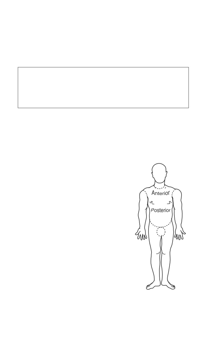
Determine the burn size based on the Rule of Nines (Fig.
28-2). A patient’s hand (palm and fingers) is approximately
1% of the total body surface area (TBSA). Only 2nd and 3rd
degree burns are included in burn size calculations.
ο Overestimation is common and may lead to over-
resuscitation and over-evacuation.
Estimate crystalloid needs for the
first 24 hours, using the following
9%
formula:
Total Volume = (2 mL) • (% burn) •
(kg weight).
18%
9%
9%
Half of this total volume is
18%
programmed for the first 8 hours
postburn, and half for the second 16
1%
hours postburn:
18% 18%
o Hourly rate, first 8 hours postburn
= (Total Volume /2) /(8h - elapsed
time in hours since burn).
Fig. 28-2. Rule of nines, showing distribution of body surface area by
anatomical part in the adult.
28.4
Burn Injuries
Assume: 40% burn, 70-kg person, time of injury 1 h ago,
(no fluids received yet).
Fluid Requirements for First 24 h = 2 x 40 x 70 = 5600 ml
One half of this to be given over first 8 h = 5600/2 = 2800 ml
But one hour has elapsed, therefore hourly rate =
2800 ml/7 h = 400 ml/h
These calculations are only an initial estimate. Patients with
inhalation injury, predominantly full-thickness burns, and
delay in resuscitation will have higher fluid requirements.
The rate of infusion of LR must be adjusted every 1-2 hours,
based on physiologic response (see below). Despite the
formula, no abrupt change is made at the 8-hour mark.
If LR is not available, use other crystalloids such as normal
saline. If crystalloid supplies are severely limited, consider
starting colloid at the 12-hour mark, at the rate recommended
for the second 24 hours (see below).
Children (< 30 kg) have a greater surface-to-weight ratio, and
their fluid requirements are greater. The formula for children
is based on 3 cc/kg/% burn.
ο In addition, children must be given a standard
maintenance infusion of D5 1⁄2NS concurrently.
Monitoring the Burn Patient
Two intravenous catheters (IVs), a Foley catheter, continuous ECG,
pulse oximeter, core thermometer, and nasogastric (NG) tube are
needed for ICU care of a patient with burns of 20% TBSA or greater.
Vital signs and fluid input/output are recorded every hour
on a flow sheet.
NG decompression is essential for all patients with burns over
20% TBSA, due to potential gastric ileus.
Secondary Survey
Perform a thorough head-to-toe secondary survey, looking
for nonthermal injuries, to include corneal abrasion, tympanic
membrane rupture, fractures, or dislocations.
If there is a question of intra-abdominal injury, diagnostic peritoneal
lavage, through burned skin if necessary, is appropriate.
28.5

Emergency War Surgery
Resuscitation Management, First 24 Hours
On an hourly basis, reassess the patient’s urine output,
which is the single most important indicator of the adequacy
of resuscitation.
Seek a urine output of 30-50 mL/h in adults or 1 mL/kg/h
in children. If the urine output is less than the target for 1-2
consecutive hours, increase the LR infusion rate by about 25%.
If it is greater than the target, decrease it by about 25%.
Avoid over-resuscitation, which may lead to edema-related
complications (eg, compartment syndromes and pulmonary
edema).
Other indices of adequate resuscitation include a decreasing
base deficit, a moderate tachycardia (typically a pulse of 100
to 130 is normal in adult burn patients), and an acceptable
mental status.
Diuretics are never indicated in the treatment of burn shock,
except when gross pigmenturia is present (see below).
Glycosuria is common following severe thermal injury and may
cause hypovolemia secondary to osmotic diuresis. Check the urine
for glucose and treat hyperglycemia with IV insulin as needed.
Resuscitation Management, Second 24 Hours
At the end of the first 24 hours postburn, discontinue the LR.
For the second 24 hours, use 5% albumin in normal saline.
The 24-hour albumin volume is as follows:
5% albumin volume = (* mL) • (% TBSA burned) • (preburn wt, kg)
% TBSA burn
30-49
50-69
70+
* mL
0.3
0.4
0.5
For example, in a burn of approximately 40% in a 70 -kg patient:
Albumin volume = (* ml) • (40%) • (70kg) = (0.3) • (2,800) =
840 mL/24 h = 35 mL/h
28.6
Burn Injuries
Burns < 30% TBSA do not require colloid infusion.
It is rarely necessary to adjust the colloid infusion rate.
If albumin is not available, fresh frozen plasma or synthetic
colloid can be used at the same dose. If none of these is
available, continue the LR until the 48th hour postburn,
monitoring urine output, and so forth.
At 24 hours, start D5W at half the last hourly rate of LR.
Follow serum sodium closely. Resuscitation is usually
complete by the 48th hour postburn. Continued evaporative
water loss replacement is needed thereafter—beware of
hypo- or hypernatremia!
Burn Wound Care
The burn wound is not an early management priority, but
must be attended to by 24 hours postburn.
Initial burn wound care includes adequate IV pain
management, removal of foreign bodies, debridement,
cleansing with surgical soap (use only saline around the
face), unroofing of all blisters, and application of a topical
antimicrobial.
Adequate wound care requires adequate pain control. Small,
intermittent boluses of IV morphine or fentanyl are effective
for background pain control. Ketamine, 1 mg/kg IV, is
generally effective for painful wound care.
Apply a topical antimicrobial cream twice daily after
thorough cleansing with a surgical detergent such as
chlorhexidine gluconate Hibiclens.
One-percent silver sulfadiazine (Silvadene), and/or 11.1%
mafenide acetate (Sulfamylon), burn creams should be
used. They are applied as a thick (1/16- to 1/8-in. thick)
layer—not as a lotion.
Following burn cream application, burns may be treated open
or closed (wrapped in gauze).
ο Extremity wounds can be wrapped in a thick layer of
roller gauze that is changed twice daily.
28.7
Emergency War Surgery
ο During the period of active wound exudation, it is helpful
to place bulky dressings beneath the burned parts to absorb
the exudate.
ο Burn cream should be reapplied to open burns as often as
needed to keep them covered.
Burn victims must be adequately immunized against
tetanus and (if arrival at the burn center will take longer
than 24 hours) should be treated with a 5-day course of
penicillin or similar antibiotic (intravenously for large
burns, orally for small ones).
Definitive burn surgery in the combat zone is generally not
recommended.
Prevent thermal (cold) stress by keeping the environment as
warm as possible (> 85°F).
Corneal abrasions in burn patients can lead to full-thickness
ulceration and blindness, and require aggressive treatment
with antibiotic ointments, preferably gentamicin or a
quinolone every 4 hours, alternating with erythromycin every
4 hours.
It is common for patients to develop a sterile, chemical
cellulitis, manifested by an erythematous rim of normal tissue
extending 1-2 cm around the wound margin. Erythema
extending beyond this margin, with other clinical evidence
of infection, likely represents beta-hemolytic streptococcal
cellulitis. Consider vancomycin if penicillin has already been
given. Treat with appropriate IV antibiotics.
Invasive gram-negative burn-wound infection is heralded by
striking changes in the color of the burn wound and a clinical
course consistent with sepsis.
ο Initiate an aminoglycoside and a semi-synthetic anti-
pseudomonal penicillin; apply Sulfamylon cream bid if
available; and plan urgent evacuation.
ο Consider subeschar clysis (injection via a spinal needle)
with the daily dose of an antipseudomonal penicillin
(ticarcillin, piperacillin) in a suitable volume of
crystalloid solution (eg, 500 mL). This is done at time of
diagnosis, and then immediately prior to excision to fascia.
28.8
Burn Injuries
Daily inspection of the burn wound by a surgeon is
essential to identify early infection complications.
Extremity Care
Carefully monitor the extremities throughout the
resuscitation period. The management of the burned
extremity can be summarized as follows:
ο Elevate;
ο Exercise burned extremities hourly;
ο Evaluate pulses and neurologic status hourly; and
ο Perform escharotomy as indicated.
In extremities with full thickness, circumferential burns,
edema formation beneath the inelastic eschar may gradually
constrict the venous outflow and, ultimately, arterial inflow.
Adequate perfusion must be assessed hourly during resuscitation.
Progressive diminution of audible arterial flow by
Doppler flowmetry is the primary indication for
escharotomy. Doppler pulses should be sought in the
palmar arch, not the wrist.
Pulses may be difficult to palpate in edematous, burned
extremities. However, in the absence of a Doppler
flowmeter, and in the appropriate clinical setting, loss of
palpable pulses may indicate a need for escharotomy.
Patients requiring escharotomy often present with a tight and
edematous extremity. They may have progressive neurologic
dysfunction such as unrelenting deep tissue pain or
paresthesias, and/or distal cyanosis.
Prior to prolonged transport, strongly consider prophylactic
escharotomy.
Note that loss of the palmar arch Doppler signal, in the presence
of adequate radial and ulnar pulses, is an indication for dorsal
hand escharotomies. These are performed over the dorsal
interossei. Digital escharotomies may be useful in some cases.
Following escharotomy, document restoration of normal
pulses and continue to monitor the patient. If one incision
fails to restore pulses, make a second incision on the other
side of the limb.
28.9
Emergency War Surgery
After escharotomy, cover wounds, including the escharotomy
incisions, in burn cream.
The patient may still develop a true intramuscular, subfascial
compartment syndrome requiring fasciotomy.
Fractures associated with thermal injury are best treated
by skeletal traction or by external fixation to permit
exposure of the burns and their treatment with topical
antimicrobials. Plaster, if used, should be bivalved
immediately to permit access for wound care and to
accommodate edema of the burned limb.
Other Considerations
Burn patients manifest a hypermetabolic state, with
hyperthermia, tachycardia, and hypercatabolism, which may
be difficult to distinguish from early sepsis.
Stress ulcer prophylaxis is critical (see Chapter 19, ICU Care).
Early enteral nutrition—once hemodynamically stable,
generally at 24 hours.
Respiratory care.
ο About one week after injury, patients with subglottic
inhalation injury may develop obstructing bloody casts.
Inhaled heparin sodium, at a dose of 10,000 units, may be
given by nebulization every 4 hours in order to prevent
the formation of these casts.
ο Subglottic inhalation injury may persist longer than
clinically evident. Extubation must be performed with
caution after adequate airway assessment.
Patients with large burns are at risk for abdominal
compartment syndrome.
Electrical Injury
High-voltage electrical injury (>1,000 volts) causes muscular
damage that often is much greater in extent than the overlying
cutaneous injury.
Examine the extremities for compartment syndrome and
perform urgent fasciotomy as needed.
Gross pigmenturia (myoglobinuria) may result, and fluid
resuscitation must be modified to protect against renal injury.
28.10
Burn Injuries
ο Pigmenturia is diagnosed by reddish-brownish urine, with
a dipstick test which is positive for blood, but with
insignificant numbers of red blood cells on microscopy.
ο Increase the hourly LR rate until a urine output of 100 ml/
h is achieved.
ο If this fails to cause a progressive clearing of the urinary
pigmenturia over 3 to 4 hours, add 12.5 g mannitol to each
liter of LR infused and consider invasive monitoring.
ο Infusion of sodium bicarbonate in water (150 mEq/L) in
order to alkalinize the urine may be useful.
Hyperkalemia may occur as a result of rhabdomyolysis, and
must be carefully assessed and treated, with calcium
gluconate infusion, insulin, and glucose.
Surgical debridement of nonviable muscle is the definitive
treatment of myoglobinuria.
High-voltage electric injury requires consideration of
deep muscle injury, with resultant rhabdomyolysis,
hyperkalemia, acute renal failure, and compartment
syndrome. Cardiac monitoring, aggressive fluid and
electrolyte management, fasciotomy, and debridement
are often required.
Patients with electrical injuries are also at high risk for spinal
fractures.
Chemical Burns
Initial treatment requires immediate removal of the offending
agent.
ο Brush any dry materials off the skin surface before copious
water lavage.
ο In the case of alkali burns, lavage may need to be continued
for several hours.
ο Resuscitate and manage just as a thermal burn.
White Phosphorus Burn
Most of the cutaneous injury resulting from phosphorus burns is
due to the ignition of clothing, and is treated as a conventional burn.
28.11
Emergency War Surgery
Fragments of this metal, which ignite upon contact with the
air, may be driven into the soft tissues.
First aid treatment of casualties with imbedded phosphorus
particles includes copious water irrigation, and placement
of a saline-soaked dressing that must be kept
continuously wet.
Profound hypocalcemia, and hyperphosphatemia, have been
described as effects of white phosphorus injury. Treat with
IV calcium.
Rapid surgical removal of the identifiable particles is often
required. UV light can be used to help locate them.
ο A dilute (1%) freshly mixed solution of copper sulfate has
been used to help identify white phosphorus particles.
However, this is no longer recommended because if the
solution is absorbed, it can cause fatal hemolysis. If it is
used, immediately wash it off with copious saline
irrigation. Never apply it as a wet dressing.
Liberally apply topical antimicrobial burn creams
postoperatively.
“How I Do It”: Excision and Grafting
Definitive burn care, including surgery and rehabilitation, is
manpower and resource intensive; therefore it is generally
inadvisable to perform excision and grafting of burns in a theater
of operations. However, under certain circumstances, this may
be unavoidable.
Patient Selection
Do not attempt to autograft patients with grossly colonized or
infected wounds. Such patients are best treated with deep
tangential excision or primary excision to fascia, followed by
immediate placement of a biologic dressing such as gamma-
irradiated allograft (Gammagraft, Promethean LifeSciences, Inc.,
Pittsburgh, PA). Many second-degree (partial thickness) wounds
are likely to heal in 14-21 days with acceptable cosmetic and
functional outcomes. Partial-thickness wounds which take longer
to heal, are likely to heal with fragile or hypertrophic scar, and
should be considered for grafting. Likewise, full-thickness burns
will only heal by contracture and should be considered for grafting.
28.12
Burn Injuries
Preparation
When performing burn surgery in a theater of operations, it
is preferable to perform several limited procedures (eg, 10%
TBSA or less at each operation) in order to limit the
physiologic stress of the operation. Plans for the operation
must be discussed and rehearsed with all personnel involved,
and the availability of OR equipment and postoperative
dressings and splints must be ascertained. At least 4 units of
PRBCs should be available for a patient undergoing a 10%
TBSA excision. A single dose of prophylactic intravenous
antibiotic, such as a first-generation cephalosporin, should
be administered. (However, antibiotics effective against
Pseudomonas and other gram-negative wound pathogens
should be considered for patients with heavily colonized
wounds.) Total IV general anesthesia (TIVA), based on
ketamine, is very effective in burn patients. Select the donor
sites to be used. Often, the anterior thighs are available and
easy to harvest. However, any area of clean unburned skin
can be harvested. Hair is removed from the donor site, and
both the area to be excised and the donor site are prepped.
Tangential vs Fascial Excision
Many surgeons recommend that extensive burn wound excision
of the extremities be performed after exsanguination and
pneumatic tourniquet application to limit blood loss. Using a
Weck knife for small areas, or a Blair knife (or similar) for large
areas, the burn is tangentially excised to the level of viable tissue.
When an area has been exsanguinated, absence of hemosiderin
staining of the dermis or fat is the usual endpoint for tangential
excision. When an area has not been exsanguinated, the usual
endpoint is diffuse punctuate bleeding (in the dermis) or viable-
appearing fat. When the surgeon believes that all nonviable
tissue in the surgical field has been excised, gauze soaked in a
1:100,000 solution of epinephrine in lactated Ringer’s can be
applied, followed by a tight ace wrap. The tourniquet is released
and the wound is reassessed after 5-10 minutes. Hemostasis in
the bed is then achieved by electrocautery. If available, topical
hemostatic agents such as spray thrombin and fibrin sealant can
be applied before letting the tourniquet down.
28.13
Emergency War Surgery
Alternatively, if the burn extends into fat and/or demonstrates
evidence of invasive burn wound infection, the burn wound
can be excised to the level of the investing muscle fascia, using
electrocautery.
Donor Site Harvesting
The subcutaneous space of the selected donor site is clysed with
a saline solution containing a 1:1,000,000 dilution of epinephrine.
This technique reduces bleeding and can be used to round out
irregular contours of the donor site when skin must be harvested
from bony or irregularly shaped areas. It is particularly
important to do this prior to harvesting the scalp to control
bleeding. It is optional for most other locations. A pneumatic
or electric dermatome is loaded with a wide blade and the
thickness of the skin to be harvested is adjusted to a depth
between 8/1,000 and 15/1,000 inch. It would be appropriate to
use 10/1,000 for grafting of most sites. Many surgeons use skin
harvested at a depth of 12/1,000 to 15/1,000 inch for the hands.
If a powered dermatome is not available, skin grafts can be
harvested using a manual dermatome or a Weck knife.
Hemostasis of the donor site is achieved with the application of
warm gauze packs, soaked in a 1:100,000 solution of
epinephrine.
At the end of the procedure, the packs are removed and the
donor site is dressed with a single sheet of rolled, fine-mesh
gauze or xeroform
(petrolatum and
3% bismuth
tribromophenate) gauze. Donor sites can alternatively be
dressed in a biosynthetic membrane material such as Biobrane.
When the donor site is small, another alternative is to apply an
occlusive transparent film dressing such as a large OpSite (Smith
& Nephew, Largo, FL) to the donor wound.
Application and Securing of the Graft
The harvested split-thickness skin may be meshed. It would be
appropriate in this case to use 1.5:1 or 2:1 for the arms; but
unmeshed skin, or skin meshed 1:1 or 1.5:1, is preferred for the
hands. If a mesher is not available, the graft can be pie-crusted
28.14
Burn Injuries
using a scalpel. The graft is applied to the prepared bed and
stapled in place. Over the hands, the graft is minimally
expanded. Bridal veil (or another product to prevent shear, such
as fine-mesh gauze) is applied over the grafted areas, followed
by a moist gauze dressing. Dressings should be kept slightly
moist, for example, by application of normal saline or aqueous
5% Sulfamylon solution (Bertek Pharmaceuticals Inc.,
Morgantown, WV) every 6-8 hours. Another option, when the
grafted area is surrounded by normal skin, is the use of a
vacuum-assisted closure device (V.A.C., Kinetic Concepts, Inc.,
San Antonio, TX).
Following dressings, the extremities are splinted, with the axilla
at 90° horizontal with bedside troughs or in an airplane splint,
elbow fully extended. Hands and wrist are splinted in the “beer
can” position: wrist slightly extended (10°), metatarsophalangeal
joints of the fingers flexed, interphalangeal joints fully extended,
and thumb in 40°-50° of abduction with interphalangeal joint
extended.
Postoperative Care
Donor sites dressed with fine-mesh gauze are treated open, with
a heat lamp applied until the gauze is dry. Grafted extremities
are immobilized for 4-5 days. Grafted sites are inspected 4-5
days after surgery. They should be inspected sooner in case of
fever, malodor, or other evidence of infection. Moist dressings
are continued until the interstices of the grafted area have
entirely closed.
Physical and occupational therapy are begun as soon as graft take
is sufficient to discontinue immobilization, usually 5 days after
surgery. Extremities are splinted in the position of function at night.
The dried gauze on the donor site is allowed to separate
spontaneously, at which time the donor site can be recropped as
necessary for further grafting. After all wounds are closed, the
patient is fitted for custom compression garments. If garments are
not available, compression can be achieved with ace bandages.
28.15
Chapter 29
Environmental Injuries
Introduction
The successful prevention and control of cold, heat, and altitude
injuries depend on vigorous command interest, the provision
of adequate clothing, and a number of individual and group
measures. The medical officer must ensure that he or she
understands how military duties impact the occurrence and
severity of environmental conditions and advise the commander
on preventive measures.
Cold Injuries
Trench foot and frostbite together have accounted for over 1 million
US casualties in WW I, WW II, and the Korean War. Influencing
factors include previous cold injury; fatigue; concomitant injury
resulting in significant blood loss or shock; geographic origin;
nutrition; tobacco use; activity; drugs and medication; alcohol;
duration and exposure; dehydration; environment (temperature,
humidity, precipitation, and wind); and clothing.
Non-Freezing Cold Injury
Chilblain.
ο Results from intermittent exposure to temperatures above
freezing, usually accompanied by high humidity and
moisture; 1 to 6 hours of exposure.
ο Swelling, tingling pain, and numbness with pink-to-red
flushing of skin (especially the fingers).
ο Extremities will be pruritic as they warm up.
ο Symptoms usually subside overnight; some superficial
scaling may occur.
ο Mild joint stiffness may occur acutely but subsides in a
few hours.
ο No permanent damage occurs.
29.1
Emergency War Surgery
Pernio.
ο Continuum of events from chilblain.
ο Exposure for > 12 hours to cold and/or wet conditions.
ο Tight-fitting footwear can shorten exposure time and
increase severity of injury.
ο Swelling is more severe; pain is more persistent than with
chilblain.
ο Thin, partial-skin thickness, and necrotic patches (from
dorsum of the hands or feet).
ο Plaques may slough without scarring but may be
particularly painful for months or years.
Trench foot.
ο
Epidemiology/clinical appearance.
♦ Occurs from prolonged exposure to cold, wet conditions
or prolonged immersion of feet at temperatures as high
as 17°C for > 12 hours. Shorter duration at or near 0°C
results in the same injury.
♦ Occurs in nonfreezing temperatures 0°C-12°C.
♦ Can occur at higher temperatures from prolonged water
immersion.
♦ Blunt trauma of marching can produce more serious injury.
♦ First symptom often is the feet becoming cold, mildly
painful, and numb.
♦ Tight footwear increases risk of trench foot.
♦ Common symptoms are “cold and numb” or “walking
on wood.”
♦ Foot may appear swollen, with the skin mildly blue,
red, or black.
♦ Limb is hot and often hyperhidrotic.
♦ On rewarming, pain is excruciating and does not
respond to pain medication, including morphine.
♦ As time progresses, liquefaction necrosis occurs distally,
but more proximal tissue may also be compromised.
♦ No sharp line of demarcation of dead from viable tissue.
♦ Nerve, muscle, and endothelial cells are most suscep-
tible to this long-term cooling.
♦ Microvascular vasospasm with tissue ischemia is the
apparent etiology of trench foot.
29.2
Environmental Injuries
♦ Postinjury sequelae include pain, numbness, loss of
proprioception, and cold feet. Hyperhidrosis with
subsequent paronychial fungal infections are common.
♦ Life-long, life-changing injury.
ο
Treatment.
♦ Prevent further cold exposure.
♦ Do not massage.
♦ Dry extremity, warm torso, and allow slow passive
rewarming of feet. Never immerse feet in warm or hot
water.
♦ Elevate feet.
♦ Rehydrate.
♦ If vesicles develop do not debride.
♦ Pain medication. The only effective approach is
amitriptyline 50-150 mg at bedtime. Other analgesics
are either completely ineffective, or (as with narcotics)
do not actually relieve pain.
♦ Blisters should be left intact; ruptured blisters require
meticulous antisepsis after unroofing.
♦ Systemic antibiotics and tetanus prophylaxis are
indicated when there are nonviable tissues, as with any
other contaminated wound, or when there is evidence
of infection.
♦ Debridement of necrotic tissue may be required in
trench foot.
♦ Macerated or damaged skin requires topical antibacter-
ial precautions.
♦ Avoid trauma.
♦ Early mobilization is vital to prevent long-term
immobility.
♦ Recovery is protracted and may require evacuation
because trench foot may lead to weeks-to-months of
pain and disability.
♦ Long-term sequelae are very common and include
sensitivity to the cold (secondary Raynaud’s phenomen-
on), chronic pain, neurological impairment, and
hyperhidrosis.
29.3
Emergency War Surgery
Frostnip.
ο Exposed skin appears red or minimally swollen.
ο Tissue is not actually damaged.
ο Not true frostbite; freezing is limited to skin surface only.
ο Signals imminent likelihood of frostbite developing.
ο Resolves quickly with warming.
Frostbite.
ο
Results from crystallization of water in the skin and
adjacent tissues exposed to temperatures below freezing.
ο
Depth and severity of injury is a function of temperature
and duration—the lower the temperature, the shorter the
time required to produce injury.
ο
At low temperatures in the presence of wind, exposed skin
can freeze within a few seconds—starts distally and
progresses up the finger or toe.
ο
Freeze-front (line where the ice is formed in the tissues) is
where liquefaction and necrosis occur. Tissues immediately
proximal to this line may also die, but therapeutic
modalities are directed at improving their survival.
ο
Clinical appearance.
♦ Skin initially becomes numb and feels stiff or woody.
♦ Mottled, bluish, yellowish, “waxy,” or “frozen.”
♦ Depth of involvement may be difficult to determine
until demarcation occurs, which may take an extended
period.
ο
Frostbite grading.
♦ Classification into degrees is primarily a retrospective
evaluation and has little treatment value.
♦ A more clinically useful grading typically divides
injuries into superficial or deep.
♦ Superficial frostbite.
◊ Involves only the skin with swelling, mild pain, and
minor joint stiffness.
◊ No blisters form.
◊ Nonmedical personnel can manage simply by
rewarming.
♦ Deep frostbite.
◊ Involves deeper tissues to include bone.
◊ White-hard, anesthetic, blanched, and inflexible.
29.4
Environmental Injuries
◊ Skin will not move over joints.
◊ On rewarming, there is great pain and a blue-gray-
to-burgundy color change.
◊ Blisters form and are clear, fluid-filled, or hemor-
rhagic (the latter indicates a more severe, deeper
injury). They should be left in place; will slough in
7-10 days without consequence.
◊ Failure to form vesicles in an obviously deep-frozen
extremity is a grave sign.
◊ Postinjury sequelae include Raynaud’s phenomenon;
pain; paraesthesias; hyperhidrosis; loss of proprio-
ception; cold, discolored feet; and gait modification.
Field treatment (first aid).
ο Superficial (blanched cheeks, nose, ears, fingertips).
♦ Warm with palm of hand or warm, wet cloth; warm
fingers in armpits.
♦ Emollients may help prevent skin from drying or
cracking.
♦ Do not massage, rub with snow, or warm part by an
open fire or high-heat source.
♦ Meticulous skin care is required.
ο Deep frostbite.
♦ Prevent further cooling of body part as well as the
patient as a whole.
♦ Apply dry, sterile bandage and elevate involved
extremity.
♦ Protect from refreezing during evacuation.
♦ Evacuate promptly to definitive medical care.
Avoid thawing and refreezing; this leads to the greatest
damage to tissue and the poorest outcome.
MTF treatment.
ο The outcome of a frozen extremity is not directly related
to length of time frozen, but more importantly to the
method of rewarming and any subsequent refreezing.
♦ If the soldier will again be at risk for refreezing, no
attempt at rewarming should be initiated; the soldier
29.5
Emergency War Surgery
should ambulate on the frozen extremities until he
reaches definitive care.
♦ For transport, the patient’s extremity should be splinted,
and padded with dry dressings and protected from heat
sources that would slowly rewarm the extremity.
ο
Rapid rewarming (without the possibility of refreezing)
is the treatment of choice.
♦ Immerse in gently circulating water (whirlpool bath) at
40°C (104°F) for at least 30 minutes longer than could
be needed to defrost all affected tissues. If deep freezing
of the leg or arm has taken place, thorough surgical
fasciotomy is mandatory prior to rewarming, to prevent
lethal increase in deep tissue pressures as ice melts.
Extremities are rewarmed until pliable and erythematous
at the most distal areas.
♦ Twice daily whirlpool baths at 40°C with topical
antibacterial added to the water, together with oral
ethanol. The alcohol reduces the need for analgesia and
may improve outcome. Other drug regimens remain
unproven.
♦ After rewarming, edema will appear within a few hours
and vesicles form within the next 6-24 hours.
♦ Intensive mobilization is essential to avoid long-term
immobility.
ο
Vesicles.
♦ Frostbite vesicles are typically left intact.
♦ Debridement is not recommended. Early surgery is only
indicated in severely infected cases. Normally surgery
should be delayed for at least 6 months.
ο
General considerations.
♦ Ibuprofen or ketorolac should be given as systemic
thromboxane/prostaglandin inhibitors.
♦ Systemic antibiotics and tetanus prophylaxis are
indicated when there are dead tissues, as with any other
contaminated wound, or when there is evidence of
infection.
♦ Dry, loose dressings may be applied.
♦ Cigarette smoking and/or nicotine use is contraindicated
during treatment due to its effect on the microvasculature.
29.6
Environmental Injuries
♦ Daily hydrotherapy is recommended. Pain control with
NSAIDs and narcotics will be needed.
♦ Sequelae include contractures, cold sensitivity, chronic
ulceration, arthritis, and hyperhidrosis.
♦ Frostbite cases will require prolonged hospital care (9 d
on average); therefore, all but those with the most trivial
injuries should be evacuated to more definitive care as
soon as possible.
♦ Early surgery is indicated only in the most severe freeze-
thaw-refreeze cases, where massive tissue destruction
has taken place, and in some more severely infected
cases. Normally, surgery should be delayed for at least
6 months (“Freeze in January, operate in July”).
Due to the inability to reliably predict the outcome in
the postthaw period, there is no role for debridement/
amputation of necrotic or potentially necrotic tissue in
the initial treatment of frostbite.
Hypothermia
Hypothermia is classically defined as whole-body cooling below
35°C. Degree of hypothermia is further defined according to
the body’s core temperature and the clinical effects seen in a
given temperature range.
Causative factors and prevention.
ο Water immersion.
ο Rain and wind.
ο Prolonged exposure to severe weather without adequate
clothing. The insulation effect of clothing is markedly
decreased with wetness, which increases the conductive
heat loss.
ο Stay dry and avoid windy exposure.
ο Shivering can provide five times the normal metabolic heat
production. Exhaustion and glycogen depletion decrease
the time of shivering. Compromise of shivering due to
inadequate food intake (skipping meals), exhaustion,
heavy exercise, alcohol, and drugs increases threat of
hypothermia.
29.7
Emergency War Surgery
Mild hypothermia > 33°C (> 91°F).
ο Shivering, hyperreflexia.
ο Amnesia, dysarthria, poor judgment, ataxia, apathy.
ο Cold diuresis.
Moderate hypothermia 28°C-33°C (82°F-91°F).
ο Standard hospital thermometers, mercury as well as
digital, cannot measure temperatures below 34°C (93°F).
ο Stupor, loss of shivering.
ο Onset of atrial fibrillation and other arrhythmias.
ο Progressive decrease in level of consciousness, respiration,
and pupillary reaction, eventual pupil dilation.
Severe hypothermia < 28°C (< 82°F).
ο Increased incidence of ventricular fibrillation, which often
occurs spontaneously.
ο Loss of motion and reflexes, areflexic at approximately
23°C (72°F).
ο Marked hypotension/bradycardia.
Profound hypothermia < 20oC (< 68°F).
ο Asystole.
ο Lowest known adult survival from accidental hypothermia
is 13.7oC (56°F).
Treatment
Prehospital (field) treatment.
ο Awake patients.
♦ Remove wet clothing; dry and insulate the patient.
♦ Give oral sugar solutions to hydrate.
♦ Walk out or transport to MTF. (This should be attempted
if it is the only alternative because it is likely to worsen
the condition.)
♦ Although walking may deepen hypothermia due to the
return of peripheral colder blood to the core, adequate
prehydration decreases the postexposure cooling.
ο Comatose patients.
♦ Patient should remain horizontal and be handled gently
to avoid inducing arrhythmias; do not massage.
♦ IV fluids, warmed to 40°C-42°C, if possible.
♦ Do not use lactated Ringer’s solution because the cold
liver cannot metabolize lactate; warm (40°C-42°C
29.8
Environmental Injuries
[104°F-107.5°F]), D5NS is the fluid of choice.
♦ Remove wet clothes, dry, insulate, and add an outer
vapor barrier. Wrap patient in multiple layers of
insulation.
♦ Limit active rewarming principally to the body’s center/
core only.
◊ Heated (40°C-45°C), humidified air/O2 is the method
of choice.
◊ Norwegian personal heater pack (charcoal heater),
with warming tube placed into insulation wrap.
◊ Forced air (Bair Hugger) with rigid chest frame.
◊ Hot water bottles in groin/axilla.
♦ Intubation and heated ventilation may be performed.
♦ If apneic and asystolic, consider CPR, because the brain
may survive longer.
Remember: The patient is not dead until he is warm and
dead. Continue resuscitation, if possible, until patient
has been rewarmed.
Medical treatment.
ο Ventilate; apply CPR if asystolic or in ventricular
fibrillation.
ο As the body cools, the peripheral vasculature constricts,
causing pooling of cold acidotic blood.
ο Rewarming the periphery of the body rather than the core
causes an inrush of this cold acidotic blood into the core,
further dropping the core temperature (afterdrop), and
worsening cardiac instability.
ο Core rewarming—peritoneal dialysis, thoracic lavage,
heated and humidified oxygen, external warm blankets,
and warm-water torso immersion.
ο For ventricular fibrillation.
♦ Bretylium tosylate, 10 mg/kg. Bretylium is the only known
effective antidefibrillation drug for hypothermia. It
remains functional in a cold heart. Other medications
have not proven effective.
♦ Warmed IV (lactate and potassium-free).
29.9
Emergency War Surgery
♦ Monitor core temperature via esophageal (preferred) or
rectal probes.
♦ Careful correction of acid/base balance.
♦ Rewarm core to 32°C (90°F) and attempt cardioversion
(360 J). Continue rewarming and repeat. Defibrillate
after every 1°C rise in temperature.
♦ Monitor potassium, glucose, temperature, and pH.
♦ Major causes of failure to resuscitate include elevating
central venous pressure too fast or too early; attempting
defibrillation when core temperature is below 32°C, or
continuing to rewarm past 33°C when potassium levels
are high and pH is low. If serum potassium levels are
high, consider the use of intravenous glucose and
insulin.
♦ Avoid other antiarrhythmics and other medications.
♦ Patients with core temperature (rectal) above 30°C can
generally be rewarmed externally in a variety of
methods including warm blankets, warm-water torso
immersion. Patients below 30°C rectal should be
considered more fragile and will often require internal
methods of rewarming (ie, warm gastric, colonic, and/
or bladder lavage; warm peritoneal lavage dialysis;
warm thoracic lavage; and arteriovenous (blood
rewarming). Lavage fluids should be warmed to 40°C-
42°C (104°F-107.5°F).
♦ Core temperature will continue to drop after the patient
is removed from the cold exposure. Continued
temperature drop can have grave prognostic implication
and increases the likelihood of fibrillation. Post-
rewarming collapse of an apparently functional heart
often leads to a nonresuscitable heart and death.
Cardiopulmonary resuscitation.
ο If cardiac monitor shows any electrical complexes, check
carefully for apical and carotid pulses before initiating
CPR. If any pulse—however thready—is present, do NOT
initiate CPR.
29.10
Environmental Injuries
Trauma patients should be considered to have hypother-
mia more profound than the core temperature indicates
and be warmed more aggressively.
Treatment of mild stable hypothermia.
ο Insulation.
ο Heat lamps.
ο Warmed IV fluids.
ο Warmed forced air (Bair Hugger). Hair dryers have been
jury-rigged for this purpose.
ο Consider arteriovenous anastomoses (AVA) warming.
♦ Immerse hand, forearms, feet, calves in water heated to
44°C-45°C (111°F-113°F).
♦ Opens AVAs in the digits causing increased flow of
warmed venous blood to the heart and decreases
afterdrop.
Treatment of severe hypothermia with hemodynamic
instability.
ο Cardiopulmonary bypass with rewarming, when available,
is the ideal technique in this circumstance because it
provides core rewarming while ensuring circulatory stability.
Heat Injury
In the military setting, heat illness occurs in otherwise healthy
individuals, and ranges from mild (heat cramps) to life
threatening (heatstroke). Individuals typically present with
exertional heat illness and are hot and sweaty, not hot and dry
as seen in classic heatstroke.
Lack of sweating is not a criterion for heatstroke. Some
military casualties of heatstroke have profuse sweating;
especially with rapid onset of heatstroke.
Minor heat illnesses include heat cramps and heat exhaustion.
Major heat injuries include exertional heat injury (EHI),
exertional rhabdomyolysis, and heat stroke. The diagnostic
categories of heat exhaustion, EHI, and heat stroke have
overlapping features and should be thought of as different
29.11
Emergency War Surgery
regions on the continuum rather than discrete disorders, each
with its own distinct pathogenesis.
Heat injury prevention.
ο
Easier to prevent than treat.
ο
Occurs most commonly in unacclimatized individuals.
♦ Acclimatization to heat requires 7-10 days.
♦ Predeployment training in artificially warm environ-
ments does aid heat acclimatization.
♦ One hour of progressively more difficult exercise sufficient
to induce moderate sweating each day will maximize
acclimatization. (Regular strenuous exercise sufficient to
stimulate sweating and increase body temperature will
result in a significant degree of heat acclimation.) Aerobic
fitness provides cardiovascular reserve to maintain the
extra cardiac output required to sustain thermoregulation,
muscular work, and vital organs in the face of heat stress.
ο
Utilize published work-rest cycle guides (eg, FM 21-10/
MCRP 4-11.1D) or work-rest cycles tailored to the
individual’s physical capacity by direct medical oversight.
ο
Water restriction/discipline leads to increased heat injury
and is contraindicated.
♦ Acclimatization does not reduce, and may actually
increase, water requirements.
♦ Service members will on average not feel thirsty until
1.5 L (1%-2%) dehydrated.
♦ Fluid intake should be monitored to ensure urine
appears dilute. Additionally, soldiers should be
monitored for body weight changes and orthostatic
blood pressure changes due to hydration.
♦ The GI tract can absorb only 1-1.5 L/h.
♦ Daily rehydration should not exceed 12 L/d orally. Too
much hydration can also be dangerous and lead to
water intoxication!
♦ Leaders must reinforce hydration by planning for all
aspects of adequate hydration—elimination as well as
consumption. (Soldiers may not drink at night to avoid
awakening and having to dress to urinate, or soldiers
may not drink prior to a convoy because no rest stops
are planned.)
29.12
Environmental Injuries
ο
MOPP gear will increase fluid losses and the incidence of
heat injuries.
ο
In the first few days of acclimatization, sweat-salt
conservation will not be fully developed. Salt depletion is
a risk if soldiers are exposed during this time to sufficient
heat or work stress to induce high sweating rates (> several
liters per day), particularly if ration consumption is
reduced. Salt depletion can be avoided by providing a salt
supplement in the form of salted water (0.05%-0.1%).
Acclimation should eventually eliminate the need for salt
supplementation
ο
Salt supplements are not routinely required and are only
recommended in rare instances where adequate rations
are not consumed.
ο
Coincidental illnesses increase heat casualty risk through
fever and dehydration. Fever reduces thermoregulatory
capacity leading to increased risk, even after clinical
evidence of illness has disappeared. Requires increased
command supervision and moderate work schedule.
ο
Sunburn and other skin diseases of hot environments reduce
the ability of the skin to thermoregulate. Sunburn must be
prevented by adequate clothing, shade, and sunscreen. Skin
diseases are best prevented by adequate hygiene.
ο
Medications that effect thermoregulatory adaptations and
increase risk of heat injury include anticholinergics,
antihistamines, diuretics, tricyclic antidepressants, major
tranquilizers, stimulants, and beta blockers.
Despite preventive measures, service members may suffer from
heat illness. One case of heat illness is a warning sign that many
others are imminent. The most life-threatening condition is
heatstroke. Severity of heat illness depends on the maximum
core temperature and duration.
Heatstroke.
Heat stroke is distinguished from heat exhaustion by the
presence of clinically significant tissue injury and/or altered
mental status. Degree of injury appears to relate to both the
degree of temperature elevation and duration of exposure.
29.13
Emergency War Surgery
ο
Clinical presentation.
♦ Heat stroke is a true emergency. Involves five organ
systems: brain, hemostatic, liver, kidneys, and muscles.
♦ Encephalopathy ranges from syncope and confusion to
seizures or coma with decerebrate rigidity. Profound
neuropsychiatric impairments present early and univer-
sally in casualties of advanced exertional heat stroke.
♦ Coagulopathy: thermal damage to endothelium,
rhabdomyolysis, and direct thermal platelet activation
causes intravascular microthrombi. Fibrinolysis is
secondarily activated. Hepatic dysfunction and thermal
injury to megakaryocytes slow the repletion of clotting
factors. Hepatic injury is common. Transaminase
enzyme elevation (values 100 or more times the upper
normal limit), clotting factor deficiencies, and jaundice
(within 24-36 h of onset). Transaminase levels may be
transient and reversible, but if they persist 48 hours, it
is indicative of more severe injury. Hypoglycemia is a
frequent complication of exertional heat stroke.
♦ Renal failure: myoglobinuria from rhabdomyolysis in
exertional heat stroke, acute tubular necrosis due to
hypoperfusion, glomerulopathy due to disseminated
intravascular coagulation (DIC), direct thermal injury,
and hyperuricemia.
♦ Muscles are often rigid and contracted: Rhabdomyolysis
is a frequent acute complication of exertional heat stroke.
Acute muscular necrosis releases large quantities of
potassium, myoglobin, phosphate, uric acid, and creatine,
and sequesters calcium in exposed contractile proteins.
If heat stroke is suspected and temperature is elevated,
cooling should not be delayed to accomplish a diagnostic
evaluation. Cooling and evaluation should proceed
simultaneously.
The patient with heat stroke requires immediate
evacuation to medical facilities with intensive care
capabilities. Active cooling should be started immediately
and continued during evacuation.
29.14
Environmental Injuries
♦ Prodromal symptoms include headache, dizziness
(lightheadedness), restlessness, weakness, ataxia,
confusion, disorientation, drowsiness, irrational or
aggressive behavior, syncope, seizures, or coma.
♦ Collapse is a universal feature of heat stroke.
♦ An individual with a core temperature of ≥ 40ºC (104°F)
and CNS dysfunction that results in delirium, convul-
sions, or coma has heat stroke.
ο
Casualties who are unconscious and have a core
temperature of ≥ 39°C (102.2°F) have heatstroke.
♦ Core temperature is often lower on arrival at a treatment
area.
♦ Seizures.
◊ Occur frequently (> 50% of cases) with heatstroke.
◊ Hinder cooling efforts.
◊ Treat with diazepam 5-10 mg.
ο
Treatment.
♦ Rapid cooling can reduce heat stroke mortality
anywhere from 50% down to 5%. Cooling by spraying
cool water over the body and vigorous fanning can be
effective though not as effective as ice water immersion.
Any effective means of cooling is acceptable.
♦ A variety of techniques have been used, and, while
evaporative cooling is less effective, the ice immersion
method may prevent safe cardiac monitoring or rapid
resuscitation.
♦ Cool water immersion (20°C) with skin massage is the
classic technique. It provides rapid cooling. Closely
monitor patient for, and prevent, shivering.
♦ Cooling with cool-water-soaked sheets or ice chips and
vigorous fanning is highly effective.
♦ Do not use alcohol in the cooling solution because
freezing of the skin can occur.
The goal of treatment is to effect a rapid lowering of the
core temperature to 38°C (101°F), without inducing
shivering.
29.15
Emergency War Surgery
♦ Rectal temperature should be closely monitored during
cooling. Discontinue cooling efforts when core temp-
erature reaches 38.3°C (101°F) to avoid hypothermia.
♦ Aspirin and acetaminophen should NOT be given to
casualties of heatstroke.
♦ Aggressive fluid resuscitation is not required. Fluid
requirements of 1 L in the first 30 minutes, with an
additional 2 L or more in the next 2 hours may be
sufficient. Because heat stroke patients are frequently
hypoglycemic, the initial fluid should include dextrose
(chilled IV fluid is of limited benefit).
◊ Base further hydration on fluid status/urinary
output (Foley required).
◊ Overhydration can lead to congestive heart failure,
cerebral edema, and pulmonary edema in the heat-
stressed lung.
♦ If shivering develops, treat with diazepam (5-10 mg IV)
or chlorpromazine (50 mg IV).
♦ Patients are frequently agitated, combative, or seizing.
Diazepam is effective for control and can be adminis-
tered IV, endotracheally, or rectally, but should be used
with caution.
♦ Airway control is essential. Vomiting is common and
endotracheal intubation should be used in any patient
with a reduced level of consciousness, or otherwise
unable to protect the airway. Supplemental oxygen
should be provided when available.
♦ Hypotensive patients who do not respond to saline
should receive inotropic support. Careful titrated use of
dopamine or dobutamine is reasonable and has the
potential added advantage of improving renal perfusion.
♦ Pulmonary artery wedge pressure monitoring should
be used in patients with persistent hemodynamic
instability.
♦ Management of encephalopathy is supportive in nature
and is directed at minimizing cerebral edema by
avoiding fluid overreplacement and by assuring
hemodynamic, thermal, and metabolic stability. IV
mannitol has been used to treat life-threatening cerebral
29.16
Environmental Injuries
edema, but is questionable unless renal function is
adequate and the patient is fully hydrated. The efficacy
of dexamethasone for treating heat-stroke-induced
cerebral edema is not known.
ο
Complications.
♦ Rhabdomyolysis and secondary renal failure due to
myoglobinuria and hyperuricemia; hyperkalemia;
hypocalcemia; and compartment syndromes due to
muscle swelling.
◊
Elevated creatine phosphokinase (CPK) (in the
thousands).
◊
Administer IV fluid and possibly furosemide to
maintain urinary output > 50 cc/h. (Assurance of
adequate renal perfusion and urine flow will
moderate the nephrotoxic effects of myoglobin and
uric acid.)
◊
Hyperkalemia can be managed by K/Na ion
exchange resin (Kayexalate) given orally or rectally
as an enema. If available, dialysis may occasionally
be indicated.
◊
Hypocalcemia does not usually require treatment.
◊
Increasing tenderness or tension in a muscle
compartment may represent increasing intracom-
partmental pressures. Direct measurement of
intramuscular pressure or fasciotomy should be
considered. Pain and paresthesia from a compart-
ment syndrome may not be present until after
permanent damage has occurred.
♦ Alkalinize urine with sodium bicarbonate IV (2 amps
NaHCO3/L D5W). Management of acute renal failure
requires exquisite attention to fluid and electrolyte
balance. Uremic metabolic acidosis and hyperkalemia
require dialysis for control.
♦ Coagulopathy due to hepatic injury.
◊ Hepatic injury is common, resulting in transaminase
enzyme elevation, clotting factor deficiencies, and
jaundice. Transaminase levels may be transient and
reversible, but if they persist 48 hours, then it is
indicative of more severe injury.
29.17
Emergency War Surgery
◊ Worst prothrombin time occurs at 48-72 hours
postinjury.
◊ Thrombocytopenia and disseminated intravascular
coagulation (DIC) peak at 18-36 hours postinjury.
◊ Beware of the coagulopathy timeframe when
planning evacuation.
◊ Subclinical coagulopathy does not require active
management. Clinically significant bleeding is an
ominous sign. Treatment is directed at reducing the
rate of coagulation and replacement of depleted
clotting factors. Intravascular coagulation can be
slowed by cautious heparin infusion (5-7 U/kg/h),
followed in 2-3 hours by FFP and platelets. Successful
management leads to a decline in indices of
fibrinolysis (eg, fibrin split products). Heparin is
tapered gradually over 2-3 days as directed by
laboratory evidence of control.
◊ Monitor for hypo- or hyperglycemia.
♦ Prognosis is worse in patients with more severe degrees
of encephalopathy. Permanent neurologic sequelae can
develop after heat stroke, including cerebellar ataxia,
paresis, seizure disorder, and cognitive dysfunction.
♦ Neurologic deterioration after initial recovery may
represent intracranial hemorrhage related to diffuse
intravascular coagulation or hematoma related to
trauma unrecognized at the time of initial presentation.
♦ Other complications include gastrointestinal bleeding,
jaundice, aspiration pneumonia, noncardiogenic
pulmonary edema, and myocardial infarction. Immune
incompetence and infection are late complications,
particularly in patients with severe renal failure.
♦ Hyperkalemia is the most life-threatening early clinical
problem. Measurement of serum potassium is an early
priority.
Heat cramps.
ο Clinical presentation.
♦ Brief, intermittent, recurring, and often excruciating
tonic muscle contractions that last 2-3 minutes.
Preceded by palpable or visible fasciculations.
29.18
Environmental Injuries
♦ Typically involve muscles of the abdomen, legs, and
arms (voluntary muscles of the trunk and extremities).
Smooth muscle, cardiac muscle, the diaphragm, and
bulbar muscles are not involved.
♦ Occur often with heat exhaustion. (Despite the salt
depletion associated with heat cramps, frank signs and
symptoms of heat exhaustion are unusual.)
♦ There are no systemic manifestations except those
attributable to pain.
♦ Occur in healthy individuals who exercise for prolonged
periods in warm environments.
♦ Occur in salt-depleted patients, generally during a
period of recovery after a period of work in the heat.
♦ Differential diagnosis: tetany due to alkalosis (hypervent-
ilation, severe gastroenteritis, cholera), hypocalcemia,
strychnine poisoning, black widow spider envenomation,
and abdominal colic.
ο
Treatment.
♦ Mild cases can be treated with oral 0.1%-0.2% salt
solutions. Salt tablets should not used as an oral salt
source.
♦ Most “sports drinks” (diluted 1:1 with water) effective
for mild cases.
♦ IV NS provides rapid relief in more severe cases
♦ Patients with heat cramps usually have substantial salt
deficits (15-30 g over 2-3 days, usual dietary intake).
These individuals should be allowed 2-3 days to
replenish salt and water deficits before returning to
work in the heat.
Heat exhaustion.
ο Clinical presentation.
♦ Thirst, headache, dyspnea, lightheadedness (orthostatic
dizziness), profound physical fatigue, anorexia,
confusion, anxiety, agitation, mood change, chills,
piloerection, nausea, and vomiting. There is no
combination of presenting symptoms and signs that is
pathognomonic.
♦ Often accompanied by heat cramps.
♦ Oliguria, clinical dehydration, ataxia, tachycardia, and
29.19
Emergency War Surgery
tachypnea resulting in symptomatic hyperventilation
with acroparesthesia and carpopedal spasm.
♦ Syncope may occur.
♦ Core temperature is < 39°C (102.2°F), even at time of
collapse.
ο
Treatment.
♦ Oral rehydration (if patient is not vomiting).
♦ Parenteral fluids produce more rapid recovery: no more
than 250 mL NS bolus without laboratory surveillance;
after 2.5 L of plain saline, add dextrose as a source of
energy (D2.51/2NaCl); subsequent fluid replacement
should be D51/2NS or D51/4NS. Individuals with
significant salt depletion have coincident potassium
depletion, often amounting to 300-400 mEq of KCl. To
begin restoration of potassium deficit, inclusion of
potassium in parenteral fluids after volume resusci-
tation is appropriate if there is no evidence of renal
insufficiency or rhabdomyolysis.
♦ Does not require active cooling; however, because
symptoms are difficult to distinguish from heat stroke,
the safest course is to provide active cooling for all
casualties who are at risk for heat stroke.
♦ Removal from hot environment.
♦ Stop exercising, move out of the sun.
Minor heat illnesses.
ο Miliaria rubra, miliaria profunda, and anhidrotic heat
exhaustion.
♦ Subacute (miliaria rubra) pruritic inflamed papulo-
vesicular skin eruption that appears in actively sweating
skin exposed to high humidity. Becomes generalized
and prolonged (miliaria profunda); lesions are truncal,
noninflamed papular, with less evidence of vesiculation
than the lesions of miliaria rubra.
♦ Each miliarial papulovesicle represents an eccrine sweat
gland whose duct is occluded at the level of the
epidermal stratum granulosum by inspissated organic
debris.
♦ Eccrine secretions accumulate in the glandular portion
of the gland and infiltrate into the surrounding dermis.
29.20
Environmental Injuries
♦ Pruritus is increased with increased sweating.
♦ Miliarial skin cannot fully participate in thermoregulatory
sweating, therefore the risk of heat illness increases in
proportion to the amount of skin surface involved.
Sweat does not appear on the surface of affected skin.
♦ Sleeplessness due to pruritus and secondary infection
of occluded glands has systemic effects that further
degrade optimal thermoregulation.
♦ Miliaria is treated by cooling and drying affected skin,
avoiding conditions that induce sweating, controlling
infection, and relieving pruritus. Eccrine gland function
recovers with desquamation of the affected epidermis,
which takes 7-10 days.
♦ Miliaria profunda causes an uncommon but disabling
disorder: anhidrotic heat exhaustion (or tropical
anhidrotic asthenia). Miliaria profunda causes a marked
inhibition of thermoregulatory sweating and heat
intolerance similar to that of ectodermal dysplasia. That
individual is more at risk for heat exhaustion and at
high risk of heat stroke in conditions tolerated by others.
♦ Evacuation to a cooler environment until restoration of
normal eccrine gland function.
ο
Heat-induced syncope.
♦ Due to a reduced effective blood volume. (Thermal
stress increases risk of classic neurally mediated
[vasovagal] syncope by aggravating peripheral pooling
of blood in dilated cutaneous vessels.)
♦ Symptoms range from light-headedness to loss of
consciousness.
♦ Typically someone standing in a hot environment.
♦ Greatest risk on first day of heat exposure, subsequent
risk decreases daily.
♦ Risk almost zero after 1 week of heat exposure; however,
syncope occurring during or after work in the heat, or
after more than 5 days of heat exposure, should be
considered evidence of heat exhaustion.
♦ Core temperature is not elevated or only very minimally so.
♦ Patient regains consciousness immediately after syncope.
29.21
Emergency War Surgery
♦ Clinical evaluation and management should be
directed toward the syncopal episode, not potential heat
illness. Treatment is oral hydration and continued
acclimatization.
ο
Heat edema.
♦ Seen early in heat exposure.
♦ Plasma volume expanding to compensate for the
increased need for thermoregulatory blood flow.
♦ In absence of other disease, condition is of no clinical
significance.
♦ Will resolve spontaneously.
♦ Diuretic therapy is not appropriate and may increase
risk of heat illness.
ο
Sunburn.
♦ Reduces thermoregulatory capacity of skin.
♦ Systemic effect: hyperthermia.
♦ Preventable.
♦ Affected soldiers should be kept from significant heat
strain until the burn has healed.
ο
Heat tetany.
♦ Rare; occurs in individuals acutely exposed to
overwhelming heat stress.
♦ Extremely severe heat stress induces hyperventilation.
♦ Manifestations include respiratory alkalosis, carpopedal
spasm, and syncope.
♦ Treatment: removal from heat source and control of
hyperventilation (rebreathing into paper bag to reverse
respiratory alkalosis).
♦ Dehydration and salt depletion are not prominent
features.
Altitude Illness
Exposure of troops to the hypobaric hypoxia of altitude results
in a decrement of performance, as well as the possible
development of altitude illness. Altitude illness spans a
spectrum from high-altitude bronchitis, to acute mountain
sickness (AMS), to death from high-altitude pulmonary edema
(HAPE) and high-altitude cerebral edema (HACE).
29.22
Environmental Injuries
Altitude basics.
The occurrence of altitude illness is based on altitude and
rapidity of ascent. Contributory factors include level of exertion,
physiologic susceptibility, age, and coexisting medical
conditions.
ο
Physiologic changes due to altitude begin to occur at just
over 1,500 m (4,900 ft).
ο
These changes are the body’s attempt to acclimatize to
altitude.
ο
Symptoms occurring below 2,250 m (7,400 ft) are rarely
due to altitude illness.
♦ Rapid ascent to high altitudes results in a high incidence
of altitude illness.
♦ Climbing Mt. Rainier brings one from sea level to 14,500
ft (4,400 m) in 36 hours and results in a 70% incidence
of altitude illness. An ascent to a similar height over
the course of 5 days would only result in a 5% incidence
of altitude illness.
♦ 10%-20% of soldiers who ascend rapidly (< 24 h) to
altitudes between 1,800 to 2,500 m (6,000-8,000 ft)
experience some mild symptoms
♦ Rapid ascent to elevations of 3,600 to 4,300 m (12,000-
14,000 ft) results in moderate symptoms in over 50% of
the soldiers, and 12%-18% may have severe symptoms.
♦ Rapid ascent to 5,300 m (17,500 ft) causes severe,
incapacitating symptoms in almost all individuals.
Descent basics.
ο Almost everything improves with prompt descent.
ο For illness requiring descent, one should try to descend at
least 1,000 m (3,300 ft) if not more.
ο A Gamow bag (USA) (portable fabric hyperbaric chamber)
or Certec SA (Europe) can temporize a patient if evacuation
/descent is not possible.
ο Symptoms typically resolve quickly with descent, but may
linger for several days.
ο Victims of HACE and HAPE should not reascend until 72
hours after symptoms abate, and then must ascend much
slower than previously.
29.23
Emergency War Surgery
ο Victims of HACE or HAPE should descend at the earliest
sign, before they become moribund and incapable of aiding
in their own descent.
ο There are no reliable predictors of susceptibility to AMS
except prior experience at altitude.
Incidence and severity of symptoms vary with initial
altitude, rate of ascent, level of exertion, and individual
susceptibility.
ο Vigorous physical activity during ascent or within 24 hours
after ascent will increase both the incidence and severity
of symptoms.
♦ If a soldier became ill previously at a given altitude he
or she will likely become ill at the same altitude unless
the ascent is slower to allow for better acclimatization.
♦ Physical fitness level has no effect on susceptibility to
altitude illness.
♦ Oral sildenafil (Viagra) 50 mg qd increases exercise
tolerance in healthy volunteers at altitude (5,200 m
[17,000 ft]), although it has not been approved for this
purpose. The role of this drug in the treatment and/or
prophylaxis of AMS and HAPE has not been established.
♦ If a rapid ascent to altitude must be made, use
prophylaxis against AMS.
Acute mountain sickness.
ο AMS is the most common form of altitude illness.
ο Onset is shortly after arrival at high altitude. Onset occurs
3-24 hours after ascent. Symptoms reach peak severity in
24-72 hours and usually subside over the course of 3-7
days.
ο Further ascent without an acclimation period usually
exacerbates symptoms and can result in increased
incidence of HAPE and HACE. The majority of AMS cases
do not progress to more serious altitude illness without
continued ascent.
ο Symptoms.
♦ Headache: Symmetric, global in location, and throbbing
in character. Most intense during night and shortly after
29.24