Emergency War Surgery (2004) - page 1

Emergency
War Surgery
THIRD UNITED STATES REVISION
2004
“All the circumstances of war surgery thus do violence to civilian
concepts of traumatic surgery. The equality of organizational and
professional management is the first basic difference. The second
is the time lag introduced by the military necessity of evacuation.
The third is the necessity for constant movement of the wounded
man, and the fourth — treatment by a number of different
surgeons at different places instead of by a single surgeon in one
place — is inherent in the third. These are all undesirable factors,
and on the surface they seem to militate against good surgical
care. Indeed, when the over-all circumstances of warfare are
added to them, they appear to make more ideal surgical treatment
impossible. Yet this was not true in the war we have just finished
fighting, nor need it ever be true. Short cuts and measures of
expediency are frequently necessary in military surgery, but
compromises with surgical adequacy are not.”
—Michael E. DeBakey, MD
Presented at Massachusetts General Hospital
Boston, October 1946
THE THIRD UNITED STATES REVISION
of
EMERGENCY WAR SURGERY
IS DEDICATED TO THE
COMBAT PHYSICIAN
Dosage Selection:
The authors and publisher have made every effort to ensure
the accuracy of dosages cited herein. However, it is the
responsibility of every practitioner to consult appropriate
information sources to ascertain correct dosages for each
clinical situation, especially for new or unfamiliar drugs and
procedures. The authors, editors, publisher, and the
Department of Defense cannot be held responsible for any
errors found in this book.
Use of Trade or Brand Names:
Use of trade or brand names in this publication is for
illustrative purposes only and does not imply endorsement
by the Department of Defense.
Neutral Language:
Unless this publication states otherwise, masculine nouns and
pronouns do not refer exclusively to men.
Library of Congress Cataloging-in-Publication Data
Emergency war surgery.-- 3rd U.S. revision.
p. ; cm.
Includes bibliographical references and index.
1. Surgery, Military--Handbooks, manuals, etc. 2. Surgical
emergencies--Handbooks, manuals, etc. 3. War wounds--
Handbooks, manuals, etc.
[DNLM: 1. Emergencies. 2. Wounds and Injuries--surgery.
3. Military Medicine. WO 800 E53 2004] I. Borden Institute
(U.S.)
RD151.E56 2004
617.9’9—dc22
2004024800
PRINTED IN THE UNITED STATES OF AMERICA
10,09,08,07,06,05
2nd printing
6,5,4,3,2
Contents
EDITORIAL & PRODUCTION
xvi
EDITORIAL BOARD
xvii
CONTRIBUTORS
xviii
ACKNOWLEDGMENTS
xx
FOREWORD
xxi
PREFACE
xxiii
PROLOGUE
xxvii
Chapter 1: Weapons Effects and Parachute Injuries
Epidemiology
1.1
Mechanism of Injury
1.2
Antipersonnel Landmines
1.6
Small Arms
1.7
Armored Vehicle Crew Casualties
1.9
Unexploded Ordnance
1.13
Parachute Injuries
1.14
Chapter 2: Levels of Medical Care
Level I
2.1
Level II
2.2
Forward Surgical Team (FST)
2.3
Level III
2.6
Level IV
2.9
Level V
2.10
Chapter 3: Triage
Categories
3.2
Special Categories
3.4
Combat Stress
3.4
Triage Decision Making
3.8
Setup, Staffing, and Operation of Triage System
3.10
Chapter 4: Aeromedical Evacuation
Medical Considerations/Requirements
4.2
vii
Medical Evacuation Precedences
4.5
Phone Numbers
4.7
Critical Care Air Transport Teams (CCATT)
4.9
Chapter 5: Airway/Breathing
Initial Management
5.1
Orotracheal Intubation
5.3
Rapid Sequence Intubation (RSI)
5.3
Difficult Airway
5.5
Surgical Cricothyrotomy
5.6
Blind Intubation
5.8
Chapter 6: Hemorrhage Control
Stop the Bleeding
6.1
Tourniquet
6.3
Internal Bleeding
6.5
Hemostatic Agents
6.6
Chapter 7: Shock and Resuscitation
Recognition and Classification
7.1
Control Bleeding
7.2
Controlled Resuscitation
7.4
Transfusion Therapy
7.6
Walking Blood Bank
7.7
Chapter 8: Vascular Access
Subclavian Vein or Internal Jugular Vein
8.1
Greater Saphenous Vein
8.3
Chapter 9: Anesthesia
Induction of General Anesthesia
9.2
Rapid Sequence Intubation
9.3
Chapter 10: Infections
Diagnosis
10.1
Common Microorganisms
10.2
viii
Treatment
10.3
Empiric Coverage
10.5
Soft Tissue Infections
10.6
Intraabdominal Infections
10.7
Pulmonary Infections
10.7
Sepsis
10.8
Dosages
10.11
Chapter 11: Critical Care
Damage Control
11.1
Resuscitation From Shock
11.2
Traumatic Brain Injury
11.3
Pulmonary System and Ventilators
11.5
Cardiovascular System
11.7
Renal System and Electrolytes
11.9
Hematologic System
11.10
Gastrointestinal System and Nutrition
11.10
Immune System and Infections
11.14
Endocrine System
11.14
Musculoskeletal System
11.15
Preparation for Evacuation
11.15
Chapter 12: Damage Control Surgery
Phases
12.1
Chapter 13: Face and Neck Injuries
Airway
13.1
Bleeding
13.2
Fracture Management
13.3
Soft Tissue Injuries
13.9
Penetrating Neck Trauma
13.12
Specific Face and Neck Injuries
13.14
Vertebral Artery
13.15
Internal Carotid Artery
13.15
ix
Internal Jugular Vein
13.15
Trachea
13.16
Esophagus
13.16
Otologic Injury
13.19
Chapter 14: Ocular Injuries
Triage
14.1
Open Globe
14.2
Anterior Segment Injuries
14.3
Cornea Chemical Injuries
14.3
Corneal Abrasions
14.4
Corneal Ulcer and Keratitis
14.5
Foreign Bodies
14.6
Hyphema
14.7
Retrobulbar Hemorrhage
14.7
Lateral Canthotomy
14.8
Orbital Floor (Blowout) Fractures
14.9
Lid Lacerations
14.10
Laser Eye Injuries
14.12
Enucleation
14.13
Chapter 15: Head Injuries
Types
15.1
Classification
15.2
Glasgow Coma Scale
15.5
Management, Medical
15.7
Management, Surgical
15.11
Chapter 16: Thoracic Injuries
Life-Threatening Injuries
16.3
Tube Thoracostomy
16.4
Resuscitative Thoracotomy
16.6
Median Sternotomy
16.9
x
Specific Injuries:
Vascular
16.11
Heart
16.12
Lung
16.12
Esophagus
16.14
Diaphragm
16.15
Chapter 17: Abdominal Injuries
Indication for Laparotomy
17.2
Diagnostic Adjuncts
17.2
Abdominal Ultrasound
17.3
Diagnostic Peritoneal Lavage
17.7
CT Scan
17.7
Wound Exploration
17.8
Stomach Injuries
17.9
Duodenum Injuries
17.9
Pancreas Injuries
17.10
Liver Injuries
17.11
Spleen Injuries
17.12
Colon Injuries
17.12
Rectum Injuries
17.14
Retroperitoneal Injuries
17.15
Abdominal Closure
17.16
Chapter 18: Genitourinary Tract Injuries
Renal Injuries
18.1
Ureteral Injuries
18.6
Bladder Injuries
18.9
Urethral Injuries
18.10
External Genitalia
18.11
Chapter 19: Gynecologic Trauma and Emergencies
Vulva Injuries
19.1
Vagina Injuries
19.2
xi
Uterus/Cervix Injuries
19.3
Emergent Total Abdominal
Hysterectomy
19.4
Adnexa
19.5
Retroperitoneal Hematoma
19.6
Gynecologic/Obstetric Emergencies
19.7
Vaginal Hemorrhage: Not Pregnant
19.7
Vaginal Hemorrhage: Pregnancy
19.8
Precipitous Vaginal Delivery
19.9
Emergency Cesarean Section
19.11
Neonatal Resuscitation
19.15
Chapter 20: Wounds and Injuries of the Spinal Column and
Cord
Classification
20.1
Pathophysiology
20.2
Transport
20.4
Cervical Spine
20.5
Halo Immobilization
20.6
Thoracic and Lumbar Spine
20.8
Emergent Surgery
20.9
Pharmacologic Treatment
20.9
General Management
20.10
Chapter 21: Pelvic Injuries
Blunt Injuries
21.1
Penetrating Injuries
21.3
Chapter 22: Soft-Tissue Injuries
Presurgical
22.1
Wound Care
22.2
Crush Syndrome
22.6
Compartment Syndrome
22.9
Fasciotomy
22.10
xii
Chapter 23: Extremity Fractures
Introduction
23.1
Transportation Casts
23.4
Shoulder/Humerus
23.8
Elbow/Forearm
23.8
External Fixation
23.10
Skeletal Traction
23.19
Chapter 24: Open-Joint Injuries
Signs
24.1
Treatment
24.2
Joint Infection
24.5
Hip Wounds
24.5
Shoulder Wounds
24.8
Chapter 25: Amputations
Indications
25.1
Technique
25.3
Postoperative Management
25.5
Transportation Casts
25.6
Chapter 26: Injuries to the Hands and Feet
Types of Injuries
26.1
Hand
26.1
Foot
26.5
Chapter 27: Vascular Injuries
Evaluation and Diagnosis
27.1
Management
27.3
Shunt Placement
27.6
Compartment Syndrome
27.8
Chapter 28: Burns
Point of Injury Care
28.1
Escharotomy
28.3
xiii
Estimation of Fluid Resuscitation
28.4
Rule of Nines
28.4
Management
28.6
Wound Care
28.7
Extremity Care, Escharotomy
28.9
Electrical Injury
28.10
Chemical Burns
28.11
Grafting: “How I Do It”
28.12
Chapter 29: Environmental Injuries
Cold Injury
29.1
Hypothermia
29.7
Heat Injury
29.11
Altitude Illness
29.22
Chapter 30: Radiological Injuries
Introduction
30.1
Triage
30.2
Decontamination
30.6
Chapter 31: Biological Warfare
Detection, Diagnosis
31.1
Decontamination
31.2
Evacuation Precautions
31.3
Bacterial Agents
31.5
Viral Agents
31.5
Toxins
31.6
Chapter 32: Chemical Injuries
Protection
32.1
Nerve Agents
32.2
Vesicants
32.3
Cyanogens
32.4
Surgical Treatment
32.6
xiv
Chapter 33: Pediatric care
Anatomic and Physiologic
Considerations
33.1
Pulmonary
33.2
Cardiovascular
33.2
Burns
33.3
Gastrointestinal
33.3
Hematology
33.4
Renal
33.4
Modified Glasgow Coma Scale
33.5
Rapid Sequence Intubation
33.6
Equipment, Age and Weight
Matched Sizes
33.7
Commonly Used Drugs
33.7
Surgical Management
33.8
Chapter 34: Care of Enemy Prisoners of War/Internees
UN and Geneva Convention
34.1
What Healthcare Providers Should Do
34.2
What Healthcare Providers Should Not Do
34.3
Recusal
34.3
Planning
34.6
Medical Photography
34.9
Security
34.10
Envoi
E.1
Appendix 1: Principles of Medical Ethics
A1.1
Appendix 2: Glasgow Coma Scale
A2.1
Appendix 3: Theater Joint Trauma Record
A3.1
Index
xxxi
xv
Editorial & Production
Borden Institute
Walter Reed Army Medical Center
Washington, DC
Andy C. Szul
Developmental/Production Editor
Lorraine B. Davis
Production Editor
Bruce G. Maston
Illustrator/Layout Editor
Douglas Wise
Layout Editor
Linette R. Sparacino
Production Editor
Jessica Shull
Illustrator
xvi
Editors
Editorial Board
Army Medical Department Center & School
Fort Sam Houston, Texas
2000-2003
David G. Burris, COL, MC, US Army
Paul J. Dougherty, LTC, MC, US Army
David C. Elliot, COL, MC, US Army
Joseph B. FitzHarris, COL, MC, US Army
Stephen P. Hetz, COL, MC, US Army
John B. Holcomb, COL, MC, US Army
Donald H. Jenkins, LTC, MC, US Air Force
Christoph Kaufmann, LTC, MC, US Army
Peter Muskat, COL, MC, US Air Force
Lawrence H. Roberts, CAPT, MC, US Navy
***
Borden Institute
Walter Reed Army Medical Center
Washington, DC
2003-2004
Dave Ed. Lounsbury, COL, MC, US Army
Matthew Brengman, MAJ, MC, US Army
Ronald F. Bellamy, COL, MC, US Army (Ret.)
xvii
Contributors
Keith Albertson, COL, MC, US Army
Rocco A. Armonda, LTC, MC, US Army
Kenneth S. Azarow, LTC, MC, US Army
Gary Benedetti, LTC, MC, FS, US Air Force
Ronald F. Bellamy, COL, US Army (Ret.)
William Beninati, LTC, MC, US Air Force
Matthew Brengman, MAJ, MC, US Army
David G. Burris, COL, MC, US Army
Frank Butler, CAPT, US Navy
Mark D. Calkins, MAJ, MC, US Army
Leopoldo C. Cancio, LTC, MC, US Army
David B. Carmacke, MAJ, MC, FS, US Air Force
Maren Chan, CPT, US Army
David J. Cohen, COL, MC, US Army
Jan A. Combs, MAJ, MC, US Army
Paul R. Cordts, COL, MC, US Army
Nicholas J. Cusolito, MAJ, NC, US Air Force
Daniel J. Donovan, LTC, MC, US Army
Paul J. Dougherty, LTC, MC, US Army
David C. Elliot, COL, MC, US Army
Martin L. Fackler, COL, MC, US Army (Ret.)
John J. Faillace, MAJ, MC, US Army
Gerald L. Farber, LTC, MC, US Army
Joseph B. FitzHarris, COL, MC, US Army
Stephen F. Flaherty, LTC, MC, US Army
Roman A. Hayda, LTC, MC, US Army
John B. Holcomb, COL, MC, US Army
Michael R. Holtel, CAPT, MC, US Navy
Stephen P. Hetz, COL, MC, US Army
Jeffrey Hrutkay, COL, MC, US Army
Annesley Jaffin, COL, MC, US Army
Donald H. Jenkins, LTC, MC, US Air Force
James Jezior, LTC, MC, US Army
Christoph Kaufmann, LTC, MC, US Army
Kimberly L. Kesling, LTC, MC, US Army
Thomas E. Knuth, COL, MC, US Army
Wilma I. Larsen, LTC, MC, US Army
xviii
George S. Lavenson, Jr., COL, MC, US Army (Ret.)
James J. Leech, COL, MC, US Army
Dave Ed. Lounsbury, COL, MC, US Army
Christian Macedonia, LTC, MC, US Army
Craig Manifold, MAJ, MC, US Air Force
Patrick Melder, MAJ, MC, US Army
Alan L. Moloff, COL, MC, US Army
Allen F. Morey, LTC, MC, US Army
Deborah Mueller, MAJ, MC, US Air Force
Peter Muskat, COL, MC, US Air Force
Mary F. Parker, LTC, MC, US Army
George Peoples, LTC, MC, US Army
Karen M. Phillips, LTC, DC, US Army
Ronald J. Place, LTC, MC, US Army
Paul Reynolds, COL, MC, US Army
Lawrence H. Roberts, CAPT, MC, US Navy
David Salas, Msgt, US Air Force (Ret.)
Joseph C. Sniezek, MAJ, MC, US Army
Scott R. Steele, CPT, MC, US Army
Allen B. Thach, COL, MC, US Army Reserve
Johnny S. Tilman, COL, MC, US Army
John M. Uhorchak, COL, MC, US Army
Steven Venticinque, MAJ, MC, US Air Force
Ian Wedmore, LTC, MC, US Army
xix
Acknowledgments
Sections of this Handbook underwent review and comment
by COL Michael Deaton (for the Surgeon General’s Integrated
Process Team on detainee medical care), LTC Glenn
Wortmann, LTC Chester Buckenmaier, LTC Peter Rhee, COL
(USAF) William Dickerson, and MAJ Clayton D. Simon.
Mr. Roy D. Flowers and Mr. Ronald E. Wallace deserve
thanks.
Although entirely unsolicited, COL Eskil Dalerius of the
Swedish Armed Forces accomplished a thorough and
insightful proofread of the Handbook, for which we are very
grateful.
The interest, efforts, and selfless service of Dr. Matthew
Brengman (formerly MAJ, MC), COL Stephen Hetz, and Dr.
Paul Dougherty (formerly LTC, MC) superseded their written
contributions, were above and beyond the call, and deserve
especial recognition.
Dave Ed. Lounsbury, MD, FACP
Colonel, MC, US Army
Director, Borden Institute
xx
Foreword
It is an honor for me to acknowledge the time, efforts, and
experience collected in this third revision of Emergency War
Surgery. Once again a team of volunteers representing the
Military Health System and numerous clinical specialties has
committed itself to delimiting state-of-the-art principles and
practices of forward trauma surgery.
War surgery, and treatment of combat casualties at far
forward locations and frequently under austere conditions,
continue to save lives. Military medical personnel provide
outstanding health support to those serving in harm’s way.
As the face of war continues to evolve, so must the practice of
medicine, to support those who so selflessly fight the global
war on terrorism. Today, American military men and women
face a new terrain of mobile urban terrorism and conflict.
Despite advances in personal and force protection provided
to our forces, they remain vulnerable to blast wounds, burns,
and multiple penetrating injuries not usually encountered in
the traditional civilian setting. This publication expertly
addresses the appropriate medical management of these and
other battle and nonbattle injuries.
The editors of this edition are to be congratulated for drawing
on the experiences of numerous colleagues recently returned
from tours of duty in Southwest Asia in order to provide as
current a handbook as possible.
I wish to publicly extend my gratitude, and that of the
American people, to the courageous men and women who
serve in the medical departments of our Armed Services. I
commend your dedicated service and acknowledge your
xxi
sacrifices, and those of your families, to provide the best
health care attainable to those who protect our nation by their
military service. I, and all Americans, are indebted to your
service.
William Winkenwerder, Jr., MD
Assistant Secretary of Defense
for Health Affairs
xxii
Preface
It is time for another revision of the Emergency War Surgery
(EWS) handbook! In addition to the fundamental advances in
the management of victims of trauma since the 1988 edition,
the format of the earlier versions was distinctly “user
unfriendly.”
This edition contains new material that updates the
management of war wounds and is filled with over 150
specially drawn illustrations. Equally important is the use of
an outline, bulleted format that is so much more concise than
the verbosity of the previous editions. Additionally, emphasis
in this edition is on the all-important “Emergency” in
Emergency War Surgery—surgery performed at levels II and III
—that constitutes the raison d’être for military surgery. Our
intent is that if given a choice of bringing a single book on a
rapid or prolonged deployment, today’s military surgeon
would choose this edition over any other trauma book.
The last revision of the Emergency War Surgery handbook was
published in 1988. Since then, world events have profoundly
affected how the US Armed Forces fight and how their
medical services provide combat casualty care. The threat of a
massive conventional war with the Soviet Union has been
replaced by a new enemy: those who espouse global
terrorism.
There are ongoing conflicts against terrorists in both Iraq and
Afghanistan, under conditions that differ radically from
Operations Desert Shield/Storm of 1990-1991. In Iraq there is
continuous urban warfare against fanatics who hide amongst
the civilian population, while in Afghanistan isolated and
sporadic but fierce small unit actions take place in
mountainous terrain. Both tactical scenarios are quite
different from what occurred in Vietnam and Operation
Desert Storm, and in what was expected for a European war
against the Soviet Union upon which the 1988 edition was
predicated.
xxiii
Military surgeons must assume a leadership role in combat
casualty care especially when faced by such changing
conditions of practice. The physicians must know what to
expect, and how to configure and prepare the team in an
austere and rapidly changing tactical environment with
available and necessary equipment. They must know how to
take care of an unfamiliar battlefield wound or injury and
manage mass casualties. Finally, they must understand the
next echelon of care, including any available capabilities, and
how to safely evacuate their patient to the higher level. This
handbook provides much of the information needed to
answer these questions.
One of the most dramatic ways in which military surgery
differs from civilian trauma management is the staged
provision of care; emergency surgery is carried out at one
locale, while definitive and reconstructive surgeries take place
at different sites. This traditional aspect of military surgery
has found new meaning in the increasing use of damage
control surgery for the most critically wounded. Here, the
initial operation is designed only to prevent further blood loss
and contamination after which resuscitation and completion
of surgery takes place, sometimes at larger, more capable
medical treatment facilities remote from the battlefield. The
US Air Force’s fielding of Critical Care Air Transport Teams
(CCATT) has revolutionized casualty care by transporting
such stabilized patients to higher levels of care during active
resuscitation. Efforts to standardize equipment across services
are in place, with the use of smaller, lightweight diagnostic
and therapeutic devices. Joint interdependence in the
treatment and evacuation of the wounded is now the
cornerstone of combat casualty care.
As a result of such advances, the Army has been able to
restructure field medical facilities essentially making them
small and mobile “building blocks.”
Despite the changes in the conditions of practice, a military
surgeon is far more likely to be deployed today than at any
xxiv
other time in our nation’s history since World War II. In the
1988 version of this handbook, BG Thomas E. Bowen quoted
Plato about the likelihood of future conflict: “Only the dead
have seen the end of war!” As military surgeons, will we be
capable and prepared to render the level of combat casualty
care befitting the sons and daughters of America? This
revision of the Emergency War Surgery handbook provides the
information needed to save the country’s and military’s most
precious resource: our soldiers, sailors, airmen, and marines.
Kevin C. Kiley, MD
Lieutenant General, Medical Corps, US Army
The Surgeon General
xxv
xxvi
Prologue
Although called the Third United States Revision, this issue of
Emergency War Surgery represents an entirely new Handbook.
Format, intent, and much of the content are new. None of the
chapters of the Second Revision has been preserved verbatim.
All material has been rewritten by new authors. Flowing
prose has been largely replaced by a bulleted manual style in
order to optimize the use of this Handbook as a rapid
reference. Illustrations are featured much more prominently
than in the earlier edition. Lastly, this text is widely available
(perhaps even more so than the printed version) electronically
on the World Wide Web and as a CD-ROM; a format neither
available nor imaginable when the second Revision was
released in 1988.
In 2000, the Surgeon General of the US Army called on the
Medical Department to revise Emergency War Surgery,
published in 1988 as the Second United States Revision and
Emergency War Surgery NATO Handbook. Responsibility for
this revision was given to the Senior Clinical Consultant in
the Directorate of Combat and Doctrine Development. He
then collaborated with the Surgeon General’s Consultant
(General Surgery) to develop a plan. These two called upon
consultants from all the Services and established an Editorial
Board of volunteers committed to a complete overhaul of the
previous Handbook. Through a series of on-line and personal
meetings coordinated by the Senior Clinical Consultant,
format and content were established. All of the chapters were
drafted and underwent review and edit by the assembled
Board at Fort Sam Houston, Texas.
Following terrorist attacks of 11 September 2001 on the
United States, US military forces were mobilized and
deployed to Afghanistan in 2001 and Iraq in 2003. The process
in place to complete this now essential Handbook was
necessarily disrupted by reassignments and deployments of
the very people who had volunteered to produce it. In lieu of
xxvii
a completed text, Borden Institute hastily published and
distributed (on-line and CD) the unedited draft manuscripts
then available as Emergency War Surgery Handbook, 2003 Draft
Version. This issue saw wide use in Southwest Asia in 2003.
In winter 2003 - 2004 Borden Institute took up the task of
completing a final version of the Handbook. With numerous
surgeons returning from yearlong tours at Combat Support
Hospitals and Forward Surgical Teams in Iraq and
Afghanistan, the decision was made to seek timely comment
on the draft manuscript. Several surgeons with fresh field
experience volunteered their subspecialty review. Substantial
updates and changes were made to many chapters including:
Anesthesia, Shock and Resuscitation, Infections, Damage
Control Surgery, Face & Neck Injuries, Extremity Fractures,
Abdominal Injuries, Burn Injuries, and Head Injuries.
At the same time material drawn from a Department of
Defense Task Force on detainee medical care (July 2004) was
adapted for the Care of Enemy Prisoners of War/Internees
chapter of this Handbook. The chapter on Triage was
expanded to include consideration of combat stress casualties.
The result of this two-stage process is this Handbook. Its
intent, and the single-minded determination of the
contributors, is the retention of lessons learned from recent, as
well as past, battlefield surgery. War surgery in the 21st
Century is not a jury-rigged art of accommodation and
compromises. Although it can include these, it is a science,
grounded on fundamentals of trauma surgery, which
recognizes as well the overriding unique principles of harsh
and austere environments, mass casualty, blast and
penetrating injury, multiple trauma, triage, staged
resuscitation, damage control surgery, time, and aeromedical
evacuation. The adage that these principles have to be
relearned by every generation of military surgeon is probably
less true than in the past. Emergency War Surgery is a
safeguard to assure this.
xxviii
Mais, plus ça change, plus c’est la meme chose. (The more things
change, the more they seem to remain the same.) Remarkable
as the enormous changes in surgical diagnostics and
therapeutics have been in the 16 years since the last edition of
the Handbook, as noteworthy - and humbling - are what
have not changed. Wound ballistics are the same and often
injuries are due to the same projectiles used 35 years ago in
Vietnam. The ghastly penetrating wounds, blast trauma, and
burns produced by present day conventional and improvised
weapons are essentially unchanged from those produced in
the last half of the 20th Century. The automatic rifle, rocket-
propelled grenade, mortar, and improvised explosive are
widely available, easy to obtain, simple to use, ferociously
lethal, and not confined to the arsenals of disciplined soldiers.
Bearers of these arms today include suicidal fanatics, women,
and children.
It is equally discouraging that although losses due to disease
have plummeted, salvage rates from severe battlefield trauma
sustained in conflict (ongoing as this Handbook goes to press)
are similar to previous wars despite improvements in armor,
surgery, critical care and evacuation. The died of wounds (%
DOW) rate during the American campaign in northwestern
Europe of 1944 - 1945 (approximately 3%) was markedly
better than that of the American Civil War (14%) nearly a
century earlier. But enormous advances in medicine and
surgery have not been reflected in substantial improvement in
lives saved in forward combat surgical facilities since then (in
World War II and Vietnam the rates were 3.5% and 3.4%
respectively).
Penetrating wounds of the head and chest are as lethal today
as they were in biblical times. Extremity fractures are still best
stabilized with external fixators, albeit a newer model.
Human blood components, with a short demanding shelf life,
have not yet been replaced despite longstanding forecasts of
synthetic products. Whole blood continues to be collected and
transfused in forward medical units as it was in the Second
xxix
World War. Bacteriologic capability to identify wound and
cavity contaminants is still unavailable in forward facilities.
Meanwhile, antibiotic resistance of numerous pathogens,
Gram negative and Gram positive, is a growing problem no
longer confined to level IV referral hospitals in the rear.
Though one can hope that major strides in these and other
areas of trauma resuscitation will be reflected in a future
edition, our more fervent hope is for mankind’s dream of
peace and the exercise of his better Angels, … whereby this
Handbook becomes altogether unnecessary.
Dave Ed. Lounsbury, MD
Colonel, Medical Corps
October, 2004
Washington, DC
xxx
Envoi
I would say that two contrary laws seem to be wrestling
with each other nowadays: The one, a law of blood and
death, ever imagining new means of destruction and
forcing nations to be constantly ready for the battlefield—
the other a law of peace, work, and health ever evolving
new means of delivering man from the scourges which
beset him. Which of these two laws will ultimately prevail
God alone knows.
—Louis Pasteur
E.1

Chapter 1
Weapons Effects
and Parachute Injuries
Just as with any medical topic, surgeons must understand the
pathophysiology of war wounds in order to best care for the patient.
Treat the wound, not the weapon.
Epidemiology of Injuries
Weapons of conventional war can be divided into explosive
munitions and small arms.
ο Explosive munitions: artillery, grenades, mortars, bombs,
and hand grenades.
ο Small arms: pistols, rifles, and machine guns.
Two major prospective epidemiological studies were
conducted during the 20th century looking at the cause of
injury as well as outcome.
ο During the Bougainville campaign of World War II, a
medical team was sent prospectively to gather data on the
injured, including the cause of injury. This campaign
involved primarily infantry soldiers and was conducted
on the South Pacific island of Bougainville during 1944.
ο US Army and Marine casualties from the Vietnam War
collected by the Wound Data and Munitions Effectiveness
Team (WDMET) in Vietnam.
US Casualties, Bougainville Campaign (WW II) and Vietnam
Weapon
Bougainville %
Vietnam %
Bullet
33.3
30
Mortar
38.8
19
Artillery
10.9
3
Grenade
12.5
11
Land mine/booby trap
1.9
17
RPG (rocket
—
12
propelled grenade)
Miscellaneous
2.6
—
1.1

Emergency War Surgery
The most common pattern of injury seen on a conventional
battlefield is the patient with multiple small fragment wounds
of the extremity.
Anatomical Distribution of Penetrating Wounds (%)
Conflict
Head and
Thorax
Abdomen
Limbs
Other
Neck
World War I
17
4
2
70
7
World War II
4
8
4
75
9
Korean War
17
7
7
67
2
Vietnam War
14
7
5
74
—
Northern Ireland
20
15
15
50
—
Falkland Islands
16
15
10
59
—
Gulf War (UK) **
6
12
11
71
(32)*
Gulf War (US)
11
8
7
56
18+
Afghanistan (US)
16
12
11
61
—
Chechnya (Russia)
24
9
4
63
—
Somalia
20
8
5
65
2
Average
15
9.5
7.4
64.6
3.5
* Buttock and back wounds and multiple fragment injuries, not
included
+
Multiple wounds
** 80% caused by fragments; range of hits 1-45, mean of 9
Mechanism of Injury
For missile injuries, there are two areas of projectile-tissue
interaction, permanent cavity and temporary cavity (Fig. 1-1).
Permanent Cavity
Sonic
Shock
Wave
Temporary Cavity
Fig. 1-1. Projectile-tissue interaction, showing components of tissue
injury.
1.2
Weapons Effects and Parachute Injuries
ο Permanent cavity. Localized area of cell necrosis,
proportional to the size of the projectile as it passes
through.
ο Temporary cavity. Transient lateral displacement of tissue,
which occurs after passage of the projectile. Elastic tissue,
such as skeletal muscle, blood vessels and skin, may be
pushed aside after passage of the projectile, but then
rebound. Inelastic tissue, such as bone or liver, may
fracture in this area.
The shock (or sonic) wave (commonly mistaken for the
temporary cavity), though measurable, has not been shown
to cause damage in tissue.
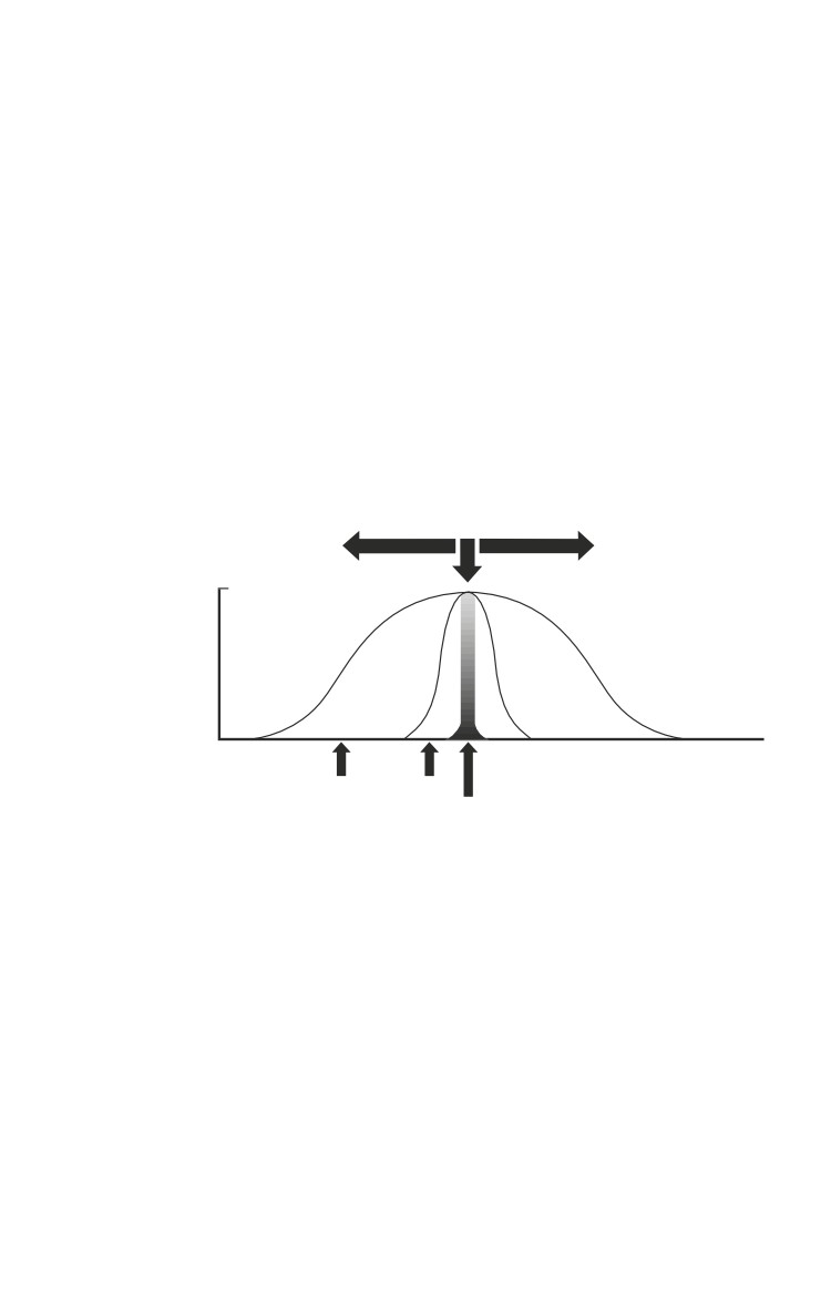
Explosive munitions have three mechanisms of injury (Fig. 1-2):
Distance From Epicenter
1.0
Probability
Of
Injury
0
Ballistic
Blast
Thermal
Fig. 1-2. The probability of sustaining a given trauma is related to the
distance from the epicenter of the detonation.
Ballistic.
ο Fragments from explosive munitions cause ballistic
injuries.
ο Fragments are most commonly produced by mortars,
artillery, and grenades.
ο Fragments produced by these weapons vary in size, shape,
composition, and initial velocity. They may vary from a
few milligrams to several grams in weight.
ο Modern explosive devices are designed to spread more
uniform fragments in a regular pattern over a given area.
1.3
Emergency War Surgery
ο Fragments from exploding munitions are smaller and
irregularly shaped when compared to bullets from small
arms.
ο Although initial fragment velocities of 5,900 ft/s (1,800 m/
s) have been reported for some of these devices, the
wounds observed in survivors indicate that striking
velocities were less than 1,900 ft/s (600 m/s). Unlike small
arms, explosive munitions cause multiple wounds.
Blast (see Fig. 1-2).
ο
The blast effects take place relatively close to the exploding
munition relative to the ballistic injury.
ο
Blast overpressure waves, or sonic shock waves, are
clinically important when a patient is close to the exploding
munition, such as a land mine.
ο
The ears are most often affected by the overpressure,
followed by lungs and the gastrointestinal (GI) tract hollow
organs. GI injuries may present 24 hours later.
ο
Injury from blast overpressure is a pressure and time
dependent function. By increasing the pressure or its
duration, the severity of injury will also increase.
ο
Thermobaric devices work by increasing the duration of a
blast wave to maximize this mechanism of injury. The
device initially explodes and puts a volatile substance into
the air (fuel vapor). A second explosion then ignites the
aerosolized material producing an explosion of long
duration. The effects from this weapon are magnified when
detonated in an enclosed space such as a bunker.
ο
Air displaced on the site after the explosion creates a blast
wind that can throw victims against solid objects, causing
blunt trauma.
Thermal.
ο Thermal effects occur as the product of combustion when
the device explodes. Patients wounded near exploding
munitions may have burns in addition to open wounds,
which may complicate the management of soft tissue injuries.
1.4
Weapons Effects and Parachute Injuries
Common Misconceptions About Missile Wounds
Misconception
Reality
Increased velocity causes
Velocity is one factor in
increased tissue damage.
wounding. An increase in
velocity does not per se
increase the amount of tissue
damage. The amount of tissue
damage in the first 12 cm of a
M-16A1 bullet wound profile
has relatively little soft tissue
disruption, similar to that of a
.22 long rifle bullet, which has
less than half the velocity.
Projectiles yaw in flight,
Unless a projectile hits an
which can create irregular
intermediate target, the
wounds.
amount of yaw in flight is
insignificant.
Exit wounds are always
This is untrue and has no
greater than entrance
bearing on surgical care.
wounds.
Full metal-jacketed bullets
The M-193 bullet of the M-16A1
do not fragment, except
rifle reliably fragments at the
in unusual circumstances.
level of the cannulure after
traversing about 12 cm of
tissue in soft tissue only.
All projectile tracts must
Elastic soft tissue
(skeletal
be fully explored, due to
muscle, blood vessels, and
the effects of the
nerves) generally heals
temporary cavity.
uneventfully and does not
require excision, provided the
blood supply remains intact.
Temporary cavity effects are
analogous to blunt trauma.
1.5
Emergency War Surgery
Antipersonnel Landmines
There are three types of conventional antipersonnel
landmines available throughout the world: static, bounding,
and horizontal spray.
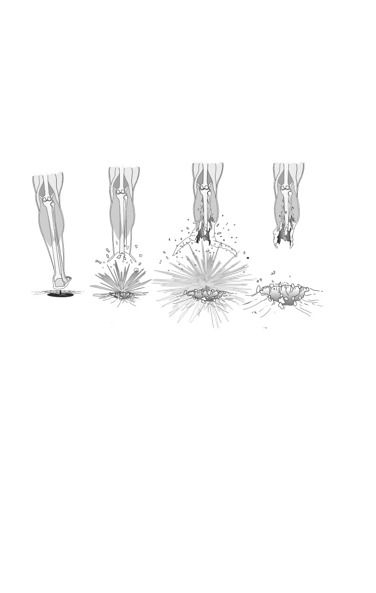
ο Static landmines are small, planted landmines (100-200 g
of explosive) that are detonated when stepped on, resulting
in two major areas of injury (Fig. 1-3).
Fig. 1-3. Mechanisms of injuries caused by antipersonnel land mines.
♦ Partial or complete traumatic amputation, most
commonly at the midfoot or distal tibia.
♦ More proximally, debris and other tissue is driven up
along fascial planes with tissue stripped from the bone.
♦ Factors influencing the degree of injury include size
and shape of the limb, point of contact with the foot,
amount of debris overlying the mine, and the type of
footwear.
ο Bounding mines propel a small explosive device to about
1-2 m of height and then explode, causing multiple small
fragment wounds to those standing nearby. These
landmine casualties have the highest reported mortality.
1.6
Weapons Effects and Parachute Injuries
ο Horizontal spray mines propel fragments in one direction.
This land mine can be command-detonated or detonated by
tripwire. The US Claymore mine fires about 700 steel spheres
of 3⁄4 g each over a 60° arc. Horizontal spray mines produce
multiple small-fragment wounds to those nearby.
An unconventional weapon (improvised explosive device,
or IED) is a fourth type of antipersonnel landmine. Either
another piece of ordnance is used, such as a grenade or a
mortar shell, or the device is completely fabricated out of
locally available materials.
Small Arms
Pistols, rifles, and machine guns.
Trends for small arms since World War II include rifles that
have increased magazine capacity, lighter bullets, and
increased muzzle velocity.
Below are some examples of the characteristics of commonly
encountered firearms seen throughout the world. The
illustrations are of the entire path of missiles fired consistently
at 5-10 m in range into ordnance gelatin tissue-simulant
blocks. Variations of range, intermediate targets such as body
armor, and body tissue will alter the wound seen.
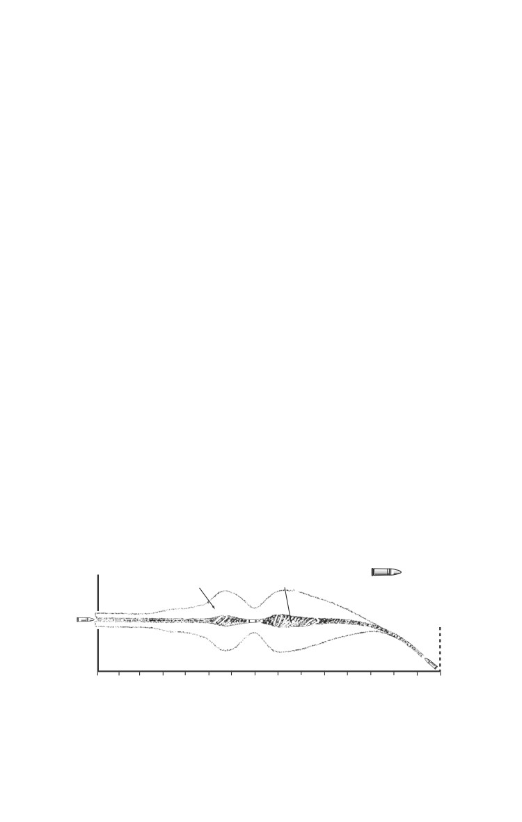
ο The AK-47 rifle is one of the most common weapons seen
throughout the world. For this particular bullet (full metal
jacketed or ball) there is a 25 cm path of relatively minimal
tissue disruption before the projectile begins to yaw. This
explains why relatively minimal tissue disruption may be
seen with some wounds (Fig. 1-4).
Temporary Cavity Permanent Cavity AK-47 7.62 mm FMC
Vel - 2340 f/s (713 m/a)
Wt - 120.5 gr (7.89 gm)
0 cm 5
10
15
20
25
30
35
40
45
50
55
60
65
70
74
Fig. 1-4. Idealized path of tissue disruption caused by an AK-47
projectile, (10% gelatin as a simulation).
1.7
Emergency War Surgery
ο The AK-74 rifle was an attempt to create a smaller caliber
assault rifle. The standard bullet does not deform in the
tissue simulant but does yaw relatively early (at about 7
cm of penetration).
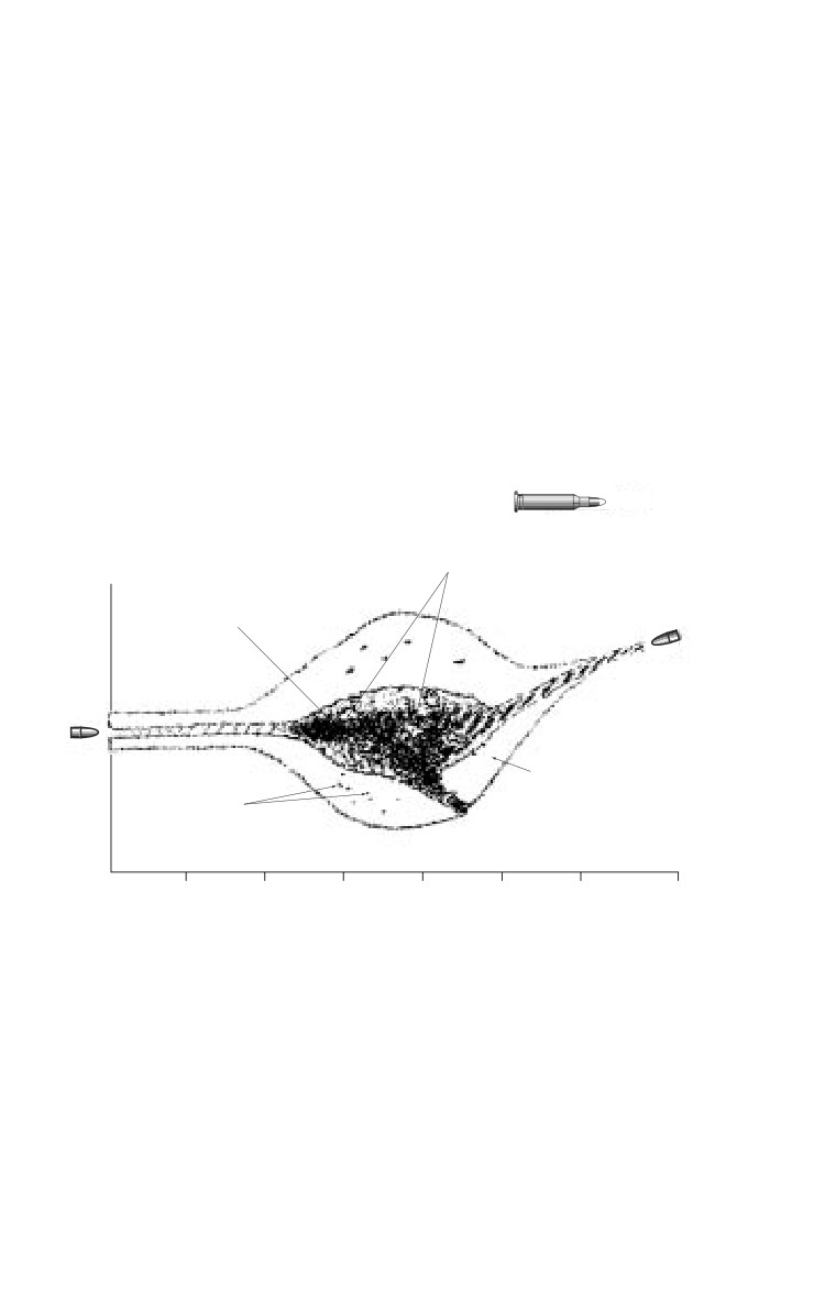
ο The M-16A1 rifle fires a 55-grain full metal-jacketed
bullet (M-193) at approximately 950 m/s. The average
point forward distance in tissue is about 12 cm, after
which it yaws to about 90°, flattens, and then breaks at
the cannalure (a groove placed around the mid section
of the bullet). The slightly heavier M-855 bullet used
with the M-16A2 rifle, shows a similar pattern to the M-
193 bullet (Fig. 1-5).
22 Cal (5.6 mm) FMC
Wt. - 55 gr (3.6)
Detached Muscles
Vel - 3094 f/s 943 m/s
Final wt - 35 gr (2.3 gm)
Permanent Cavity
36% Fragmentation
Temporary Cavity
Bullet
Fragments
5
10
15
20
25
30
35
Fig. 1-5. Idealized path of tissue disruption caused by an M-193 bullet
fired from the M-16A1 rifle (10% gelatin as a simulation).
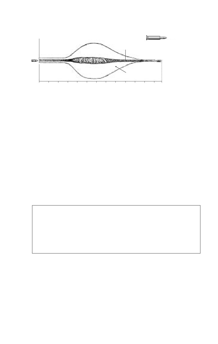
ο The 7.62 mm NATO rifle cartridge is still used in sniper
rifles and machine guns. After about 16 cm of penetration,
this bullet yaws through 90° and then travels base forward.
A large temporary cavity is formed and occurs at the point
of maximum yaw (Fig. 1-6).
1.8
Weapons Effects and Parachute Injuries
Permanent
7.62 mm NATO
Cavity
Vel - 2830 f/s (862 m/a)
Wt - 150 gr (9.7 gm) FMC
Temporary
Cavity
0 cm 5
10
15
20
25
30
35
40
45
50
55
60 65
Fig. 1-6. Idealized path of tissue disruption caused by the 7.62 mm
projectile, (10% gelatin as a simulation).
Armored Vehicle Crew Casualties
Since the first large scale use of tanks during WWI, injuries
to those associated with armored vehicles in battle have been
a distinct subset of combat casualties.
Tanks, infantry fighting vehicles, armored personnel carriers,
armored support vehicles, and “light armored vehicles.”
ο Light armored vehicles tend to use wheels rather than
tracks for moving and have lighter armor. The main
advantage for these vehicles is to allow for greater mobility.
Compared to infantry, injuries to those inside or around
armored vehicles are characterized by:
Decreased overall frequency.
Increased severity of injury and mortality (up to 50%).
Increased incidence of burns and traumatic
amputations.
There are three main types of antiarmor weapons on the
battlefield today.
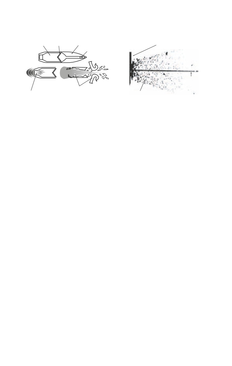
ο Shaped charge (Fig. 1-7a).
♦ The shaped charge or high explosive antitank (HEAT)
round consists of explosives packed around a reverse cone
of metal called a melt sheet or a liner. This is the principle
behind the warhead of the RPG.
1.9
Emergency War Surgery
a
b
Explosive Melt Sheet
Nose Cone
Target Material
Fuse
Armor
Jet
Gas and Fragments of
Exploding Charge
Melt Sheet
Spall Material
Fig. 1-7. (a) Disruptive mechanisms of the shaped-charge warhead,
(b) diagram taken from photograph of an actual detonation of shaped-
charged warhead against armor plate caused by antitank land mines.
♦ Shaped charges range in diameter from the 85 mm
RPG-7 to the 6-in diameter tube launched, optically
tracked, wire guided (TOW) missile.
♦ If the armor is defeated by the shaped charge, there
are two areas of behind-armor debris.
◊ First, there is the jet of the shaped charge itself. This may
cause catastrophic wounds to soldiers who are hit, or it
may ignite fuel, ammunition, or hydraulic fluid.
◊ There is a second type of debris, called spall, which
is material knocked off from the inside face of the
armored plate. This produces a spray of small,
irregularly shaped fragments inside of the
compartment (Fig. 1-7b).
ο
Kinetic energy round.
♦ The kinetic energy
(KE) round contains an
aerodynamically shaped piece of hard metal (such as
depleted uranium or tungsten) shaped like a dart. The
metal is usually encased in a carrier or sabot that falls
away from the projectile after it leaves the barrel.
Fragments of depleted uranium should be treated during
initial wound surgery as any retained metal foreign body
should. There is a hypothetical risk, over years, that
casualties with retained depleted uranium fragments may
develop heavy metal poisoning. This concern by itself
does not justify extensive operations to remove such
fragments during initial wound surgery.
1.10
Weapons Effects and Parachute Injuries
♦ Injuries to those inside a vehicle are due to the direct
effects of the penetrator or from fragments knocked
off the inside face of the armored plate. The range of
fragment masses may be from a few milligrams to over
a kilogram.
ο
Antitank landmines.
♦ Blast mines are those with a large explosive filler of 4-
5 kg. Injuries are often from blunt trauma due to
crewmembers being thrown around inside the vehicle
after it detonates the mine.
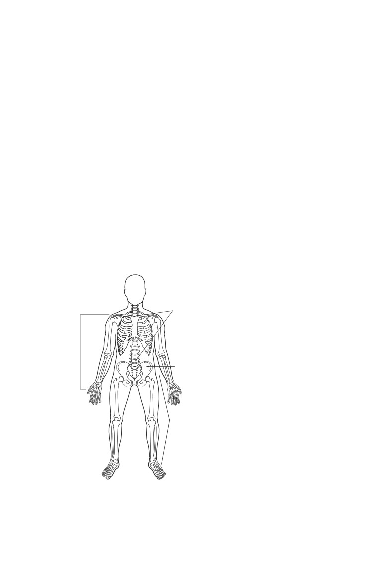
♦ Closed fractures of the upper and lower extremities and
spine are common (Fig. 1-8).
♦ A modification of the shaped charge is the Miznay-
Schardin antitank mine that creates a projectile or large
metal slug to cause damage to the vehicle. This is less
likely than a conventionally shaped charge to be broken
up by intermediate targets.
20.1%
13.5%
Fig. 1-8. Distribution of fracture
sites sustained within an
armored vehicle that had
detonated a land mine (Soviet
2.8%
data from Afghanistan, early
1980s).
63.6%
1.11
//////////////////////////////////////////